Tag: Hippocampus

  • Study identifies DeltaFosB in the hippocampus as a key driver of cocaine relapse, opening a path to targeted treatments

    Study identifies DeltaFosB in the hippocampus as a key driver of cocaine relapse, opening a path to targeted treatments

    Scientists at Michigan State University have pinpointed a brain protein that appears to be essential for the circuit changes that fuel cocaine relapse, offering a clearer biological explanation for why cravings can persist long after use stops. The findings, published in Science Advances and supported by the US National Institutes of Health, focus on a molecule called DeltaFosB.

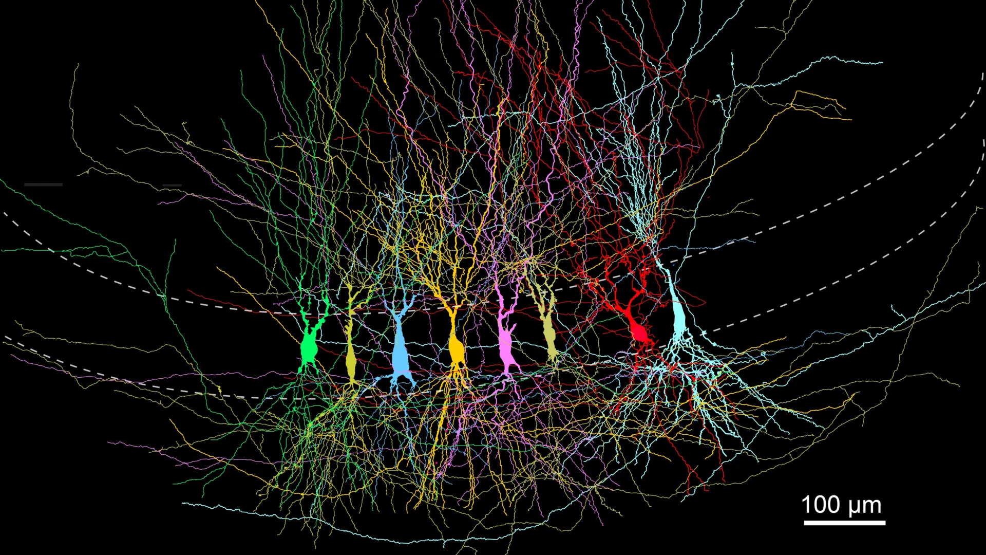
    The research highlights the hippocampus, a region central to memory and learning, and its interaction with reward pathways involved in drug seeking. By linking relapse risk to durable changes in these circuits, the study adds weight to the view that cocaine addiction is driven by brain biology rather than willpower alone.

    How cocaine rewires memory circuits

    Unlike opioids, stopping cocaine does not typically cause severe physical withdrawal, yet relapse remains common, and no FDA-approved medication is specifically indicated for cocaine use disorder. Cocaine’s surge of dopamine reinforces drug-taking, while memory-linked cues can later reignite the urge to use.

    Using mouse models and a specialized CRISPR-based approach, the team found that DeltaFosB acts like a genetic switch in a pathway connecting reward centers and the hippocampus. With repeated cocaine exposure, DeltaFosB accumulates and changes how neurons respond, increasing the drive to seek the drug.

    Genes that intensify cocaine seeking

    The researchers also identified genes influenced by DeltaFosB after longer-term cocaine exposure, including calreticulin, which helps regulate how neurons communicate. In experiments, higher calreticulin activity appeared to boost signaling in pathways that promote continued cocaine seeking.

    Crucially, the study suggests DeltaFosB is not merely associated with these adaptations but required for them to fully develop. When the protein’s role was disrupted, cocaine did not produce the same patterns of brain activity changes linked to persistent drug seeking.

    What this could mean for treatment

    Because many of the implicated genes and circuits are conserved across mammals, the authors say the findings could help guide human research, though direct clinical implications remain years away. The group is now collaborating with the University of Texas Medical Branch to develop compounds aimed at altering how DeltaFosB binds to DNA.

    Future work will also explore how hormones may shape these circuits and whether addiction-related brain adaptations differ between males and females. Such insights could eventually support more personalized approaches to treating cocaine use disorder.

  • Study of the hippocampus suggests newborn brains start densely wired, then prune connections for sharper memory

    Study of the hippocampus suggests newborn brains start densely wired, then prune connections for sharper memory

    The hippocampus, a brain region essential for forming memories and mapping space, may develop in a way that challenges the long-held idea of the mind as a blank slate. New research from the Institute of Science and Technology Austria suggests key memory circuits begin life with unusually dense wiring that is later trimmed and refined.

    In a study published in Nature Communications, scientists examined how a major hippocampal network changes after birth in mice. The team focused on CA3 pyramidal neurons, cells widely seen as central to storing and retrieving memories.

    How the CA3 circuit develops

    Using patch-clamp recordings and high-resolution imaging, researchers compared the CA3 network across early postnatal stages, adolescence, and adulthood. The methods allowed them to measure tiny electrical signals and map how strongly neurons were connected at different ages.

    The results pointed to an early-life circuit that is highly connected and seemingly random, followed by a gradual shift toward fewer but more organized links. Rather than adding connections over time, the network became more efficient by losing many of its initial ones.

    Pruning may boost memory efficiency

    Lead researcher Peter Jonas said the pattern fits a pruning model in which the system starts full and then becomes streamlined. The researchers argue that an initially exuberant network could help the hippocampus quickly integrate different kinds of sensory information into usable memories.

    If the brain started with far fewer built-in links, the team notes, neurons would first need more time to find and connect to each other, potentially slowing early information processing. The study adds to broader evidence that brain development often involves overproduction of connections followed by activity-dependent pruning.

    While the work was conducted in mice, the hippocampus is highly conserved across mammals, making the findings relevant to ongoing debates about how genetics and experience shape learning. The authors say future research will need to clarify what signals drive which connections are kept or removed, and how this process relates to memory performance.

  • UCSF study flags FTL1 protein as a driver of brain aging, and a potential new target to restore memory

    UCSF study flags FTL1 protein as a driver of brain aging, and a potential new target to restore memory

    Aging can take a heavy toll on the hippocampus, the brain region central to learning and memory. Researchers at the University of California, San Francisco report they have identified a protein that may play an outsized role in that decline.

    In a study published in Nature Aging, the team points to FTL1, a ferritin-related protein involved in iron handling, as a key molecular change seen in older mouse hippocampus. The researchers say shifting FTL1 levels altered memory performance and the strength of neural connections in ways that tracked with age.

    A standout signal in aging brains

    To pinpoint what changes over time, scientists compared gene and protein patterns in the hippocampus of young and older mice. Among many measurements, FTL1 emerged as the most consistent difference between age groups.

    Older mice had higher FTL1 levels alongside fewer synaptic connections and worse results on cognitive tasks. The authors report that the pattern suggested more than a passive marker of aging, raising the possibility that FTL1 helps drive the process.

    What happened when FTL1 was altered

    When researchers increased FTL1 in young mice, the animals developed brain and behavioral changes resembling those seen in older mice. The hippocampus showed reduced connectivity, and performance on memory-related testing declined.

    Cell experiments offered a potential explanation: neurons pushed to make more FTL1 formed simpler structures, with fewer branching extensions needed for complex signaling. That shift, the team argues, could help explain how elevated FTL1 weakens hippocampal circuitry.

    Can memory decline be reversed?

    In older mice, lowering FTL1 was linked to improved synaptic connections and better memory test performance. Senior author Saul Villeda said, “It is truly a reversal of impairments,” emphasizing that the effect went beyond delaying decline.

    The group also reported a metabolic component, with higher FTL1 associated with slower energy use in hippocampal cells. In lab settings, boosting cellular metabolism with an experimental compound reduced the harmful effects tied to elevated FTL1, pointing to possible therapeutic angles.

    Experts caution that mouse findings do not automatically translate to human brain aging, and any treatment approach would require extensive safety and efficacy testing. Still, the UCSF team argues that targeting FTL1 or related metabolic pathways could eventually open a new route for interventions aimed at age-related cognitive decline.