Tag: PET skenavimas

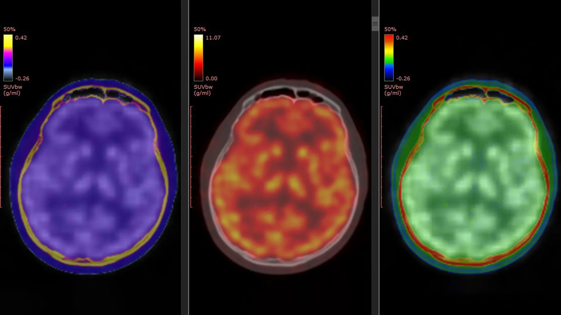
  • PET brain scans map ketamine’s rapid antidepressant effect, pointing to a potential biomarker for treatment-resistant depression

    PET brain scans map ketamine’s rapid antidepressant effect, pointing to a potential biomarker for treatment-resistant depression

    Researchers in Japan have reported some of the clearest human evidence yet of how ketamine can relieve symptoms of treatment-resistant depression, using PET brain imaging to track changes in key glutamate receptors. The work adds molecular detail to a treatment already known for acting faster than standard antidepressants.

    Major depressive disorder is a leading cause of disability worldwide, and a substantial share of patients do not improve after trying multiple first-line therapies. For those with treatment-resistant depression, ketamine and the related medicine esketamine have drawn attention because some patients feel relief within hours or days rather than weeks.

    What the PET scans measured

    The study, published in Molecular Psychiatry, used a PET tracer called [11C]K-2 designed to visualize AMPA receptors, proteins that help regulate communication between brain cells. Scientists have long suspected that AMPA receptor activity is central to ketamine’s antidepressant effects, but direct confirmation in living people has been limited.

    The researchers combined data from three clinical trials, comparing 34 patients with treatment-resistant depression against 49 healthy participants. Patients received intravenous ketamine or placebo over a two-week period, with PET scans taken before treatment and after the final infusion.

    Receptor shifts tied to symptom relief

    Before treatment, the PET data suggested patients with treatment-resistant depression had region-specific differences in AMPA receptor availability compared with healthy controls. After ketamine, the brain changes were not uniform, instead appearing as shifts in particular areas involved in mood and reward processing.

    Crucially, the degree and location of AMPA receptor changes tracked with how much a patient’s depressive symptoms improved. The authors highlighted especially notable shifts in regions linked in prior research to depression circuitry, arguing the images provide a direct bridge between earlier animal findings and human clinical response.

    Why it could matter clinically

    If replicated, AMPA receptor PET imaging could become a candidate biomarker to help predict who is most likely to benefit from ketamine, and to guide dosing or treatment strategies. That could be valuable because ketamine response can vary, and clinicians are seeking ways to personalize care while balancing benefit, side effects, and monitoring needs.

    The researchers caution that PET imaging is complex and not widely available, and larger studies would be needed before it could influence routine practice. Still, mapping ketamine’s effects at the receptor level may also support development of new rapid-acting antidepressants that target similar pathways with fewer practical barriers.

  • Loss of Smell May Signal Early Alzheimer’s: New Study Points to an Immune Trigger

    Loss of Smell May Signal Early Alzheimer’s: New Study Points to an Immune Trigger

    A subtle decline in the sense of smell could be among the earliest detectable changes linked to Alzheimer’s disease, potentially emerging well before clear memory problems. New research from Germany suggests the shift may be driven by the brain’s immune cells damaging key odor-processing connections.

    The study, led by scientists at the German Center for Neurodegenerative Diseases (DZNE) and Ludwig Maximilian University of Munich, focuses on microglia, immune cells that help maintain brain health. Researchers report that in early Alzheimer’s, microglia may begin dismantling nerve fibers needed for normal smell perception.

    How the brain’s smell circuit changes

    The team examined communication between the olfactory bulb, which processes odor signals, and the locus coeruleus, a brainstem region involved in sensory regulation and other core functions. Long nerve fibers from the locus coeruleus help tune activity in the olfactory bulb, supporting normal smell processing.

    According to the researchers, early Alzheimer’s-related alterations make these fibers appear abnormal to microglia. In response, microglia break down the connections, which could help explain why smell deficits can appear early in the disease course.

    An eat-me signal on neurons

    The study points to changes in the nerve fiber membrane as a likely trigger. A molecule called phosphatidylserine, typically kept on the inner side of the cell membrane, was observed on the outside, where it can act as an immune cue.

    Microglia are known to respond to this kind of signal during normal synaptic pruning, a process that removes unused or impaired connections. The researchers suggest that in early Alzheimer’s, abnormal neuron activity may prompt this membrane shift, leading microglia to remove fibers that are still needed.

    Evidence from mice, tissue, and PET scans

    To support the mechanism, the scientists combined results from Alzheimer’s-like mouse models with analyses of human brain tissue and PET imaging data from people diagnosed with Alzheimer’s or mild cognitive impairment. Together, these lines of evidence point to immune-driven damage occurring at an early stage.

    The findings also connect to a growing push for earlier diagnosis, as newer Alzheimer’s treatments are generally aimed at earlier phases of the disease. Researchers say a better understanding of smell-related changes could help identify people who should receive further testing before cognitive symptoms become pronounced.

    Smell loss can have many causes, including aging, infections, allergies, and other neurological conditions, so it is not a stand-alone diagnostic sign. Still, the study strengthens the case that changes in olfaction may offer a practical early clue worth taking seriously in Alzheimer’s research and clinical follow-up.