Tag: Prefrontal cortex

  • NYU study maps a prefrontal naming network, offering new clues to why word retrieval can fail

    Scientists at New York University have mapped a brain network linked to naming and word retrieval, a core function that can break down after stroke, traumatic brain injury, or neurodegenerative disease. The work helps explain why some people can name an object they see but struggle to find words in everyday conversation.

    The study, published in Cell Reports, points to a left-lateralized network involving the dorsolateral prefrontal cortex and nearby frontal regions. Researchers say the findings refine how neuroscience understands the step-by-step process of turning meaning into spoken words.

    How researchers mapped naming circuits

    The team analyzed electrocorticography recordings, a method that measures brain activity directly from the cortical surface during clinical monitoring. Data came from 48 neurosurgical patients, allowing unusually precise timing and localization of language-related signals.

    Using computational clustering, the researchers identified two partially overlapping systems involved in naming. One system tracked semantic processing, linking words to meaning and responding to how expected a word was within a sentence.

    Auditory naming highlights dorsal hub

    A second system was tied to articulatory planning and speech production, showing activity patterns that were less dependent on whether words were presented visually or through sound. This network was centered more ventrally in frontal and precentral regions associated with speech motor planning.

    The results also revealed a ventral-to-dorsal gradient across the prefrontal cortex, with a dorsal frontal area emerging as a key hub for mapping sounds to meaning in auditory contexts. The authors argue this dorsal prefrontal contribution has been underappreciated in earlier models.

    Why the findings matter clinically

    Clinicians frequently see anomia, the difficulty of retrieving words, in patients with focal brain damage and in conditions such as primary progressive aphasia. By separating semantic integration from articulatory planning, the study may help guide more targeted assessments and rehabilitation strategies.

    The work could also inform brain-computer interface research aimed at restoring communication, by clarifying which neural signals best reflect the intent to name a concept. While the authors caution that translation to devices and therapies will take time, the map provides a clearer target for future studies.
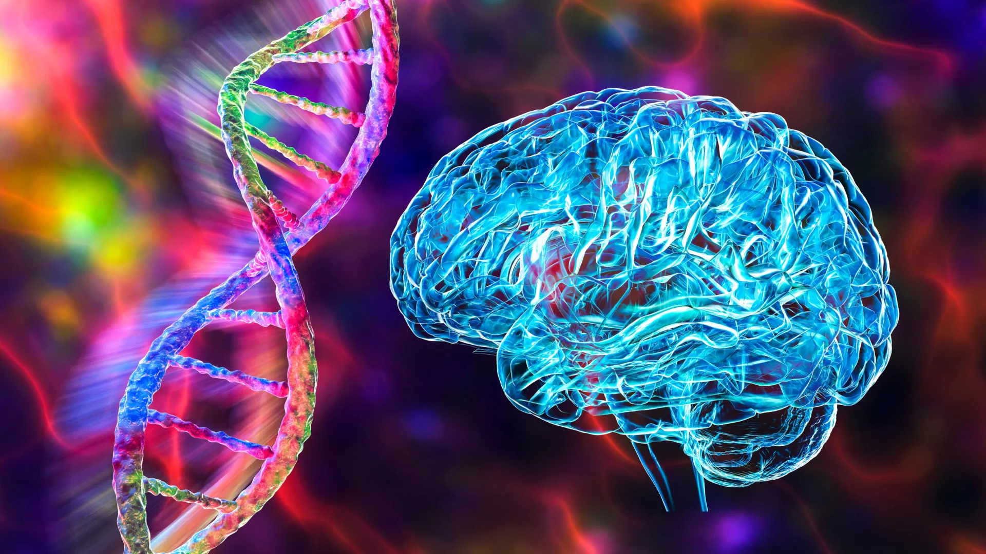
  • MIT study links GRIN2A mutation to slower reality updating in schizophrenia, pointing to a treatable brain circuit

    MIT study links GRIN2A mutation to slower reality updating in schizophrenia, pointing to a treatable brain circuit

    Researchers at MIT report that a mutation in the gene GRIN2A may interfere with how the brain updates beliefs when new information arrives, a cognitive difficulty often seen in schizophrenia. In mouse experiments, the change was tied to slower, less adaptive decision-making in a shifting environment.

    Schizophrenia affects about 1% of people and has a strong genetic component, though the biology connecting risk genes to symptoms has been hard to pin down. Large genomic studies have identified many associated variants, but many sit in non-coding DNA, making their functional impact difficult to interpret.

    From genetic signal to mechanism

    To narrow in on mutations that directly alter proteins, the team drew on large-scale exome sequencing that compares protein-coding regions across people with schizophrenia and unaffected controls. That work has helped highlight a smaller set of genes where rare disruptive mutations can substantially increase risk.

    GRIN2A stands out because it encodes a subunit of the NMDA receptor, a key component of glutamatergic signaling involved in learning, plasticity and cognitive control. NMDA receptor dysfunction has long been considered relevant to schizophrenia, but linking specific mutations to circuit-level effects has remained challenging.

    Decision task reveals slower adaptation

    In the study’s behavioral task, mice chose between two levers with different reward sizes and different effort costs that changed over time. Typical mice shifted to the more efficient option once the higher-reward choice became too costly, reflecting flexible updating as conditions evolved.

    Mice carrying the GRIN2A-related mutation took longer to commit, continuing to alternate between choices after the balance of effort and reward had effectively changed. The researchers interpret the pattern as reduced ability to incorporate new evidence quickly, leaving prior expectations to dominate behavior for longer.

    A circuit that can be nudged

    Brain measurements pointed to altered activity in the mediodorsal thalamus and its connections with the prefrontal cortex, a pathway central to executive function and decision-making. The team reports that this thalamocortical circuit appeared to represent changing option values differently in the mutated mice.

    Using optogenetics to activate neurons in the mediodorsal thalamus, the researchers were able to push behavior toward the more adaptive pattern seen in control animals. While only a subset of patients would be expected to carry GRIN2A mutations, the results suggest the same circuit could contribute to cognitive symptoms across broader groups.

    The authors frame the work as a step toward treatments that target cognition, an area where many patients continue to experience impairment even when hallucinations or delusions are reduced. Next efforts focus on identifying druggable nodes in the thalamus–prefrontal pathway that might restore more flexible updating without invasive methods.