Tag: UC Berkeley

  • UC Berkeley study points to oxytocin as the fast track to friendship, and why some bonds take longer to form

    UC Berkeley study points to oxytocin as the fast track to friendship, and why some bonds take longer to form

    New research from the University of California, Berkeley suggests the hormone oxytocin helps speed up the early stages of friendship formation, sharpening the sense of preferring a familiar peer over a stranger. The work, published in Current Biology, adds nuance to oxytocin’s popular image by focusing on how quickly and selectively social bonds take shape.

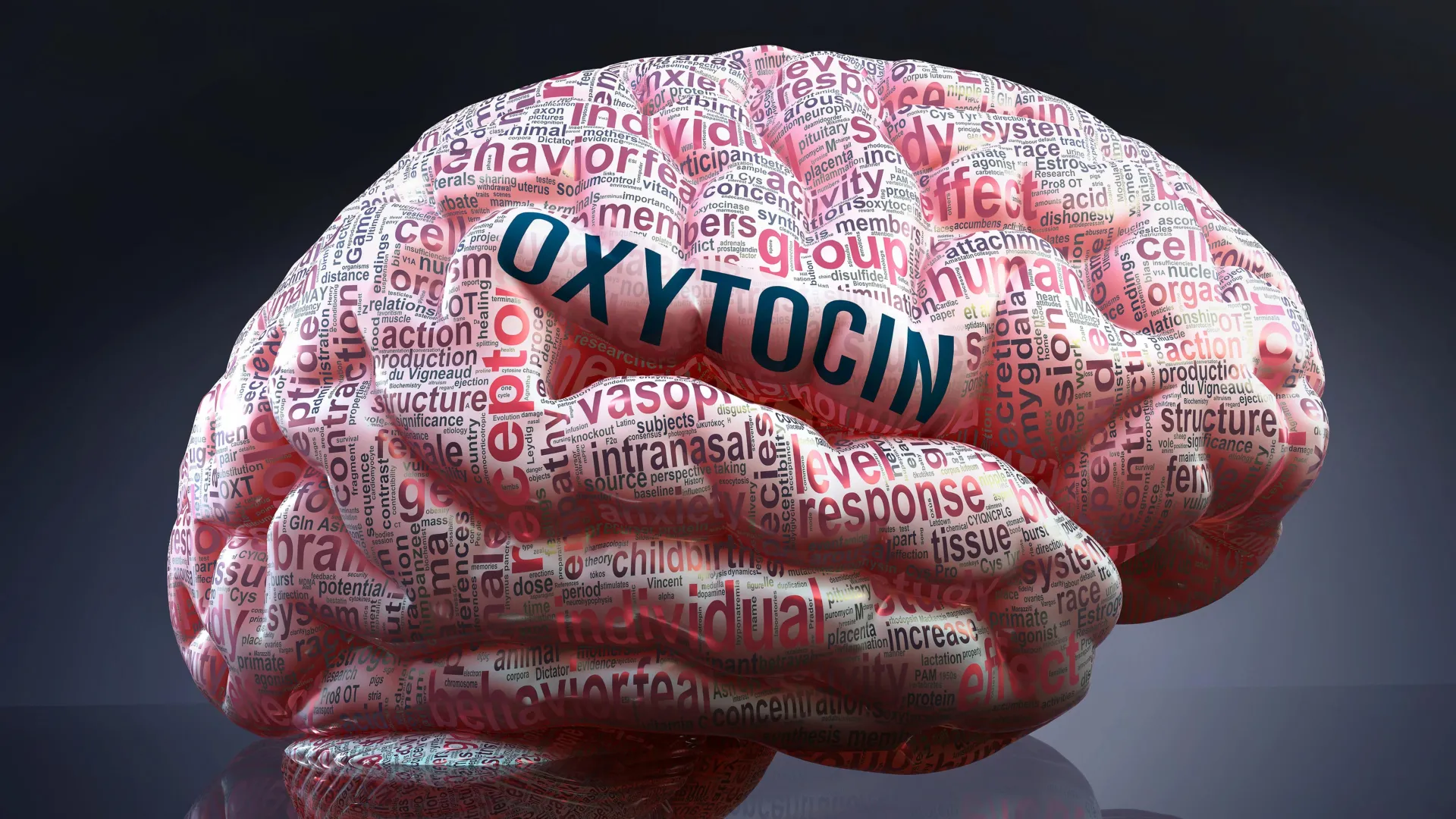
    Oxytocin is released during a range of social and bodily experiences, including touch, sex, childbirth and breastfeeding, and it acts in the brain as a neuromodulator. While it is often linked with closeness and trust, scientists have also associated oxytocin signaling with social defensiveness, including stronger in-group and out-group behavior.

    The team studied prairie voles, a species widely used to examine social bonding because individuals form stable, selective relationships. Instead of focusing only on mating pairs, the researchers emphasized peer bonds that resemble human friendships, such as choosing to huddle and groom with one familiar partner rather than spending time with strangers.

    What changed without oxytocin receptors

    Using prairie voles engineered to lack oxytocin receptors, the researchers found the animals were slower to form a peer preference. In tests where typical voles show a strong preference after about 24 hours, the receptor-deficient animals often needed up to a week to reliably choose a familiar partner.

    The difference was not simply that the animals became less social overall. The findings point to reduced selectivity, meaning the altered animals were less consistent about who they sought out and were quicker to lose track of established partners when placed into new group settings.

    Friendship selectivity, not just sociability

    In a mixed-group, multi-room setup designed to mimic a party-like environment, typical voles spent early time near known companions before gradually mingling. Voles without oxytocin receptors mixed more freely from the start, behaving as if prior peer connections carried less weight.

    In another test measuring social motivation, female voles usually worked harder to access a familiar peer than a stranger. The receptor-deficient animals still showed motivation for a mate, but not for a friend, indicating that oxytocin signaling may matter more for the reward value of peer bonds than for mating bonds.

    A new look with oxytocin nanosensors

    To examine whether the brain compensated for missing receptors by releasing more oxytocin, researchers used an oxytocin nanosensor that fluoresces when it detects the molecule. Measurements indicated no excess oxytocin release and, instead, lower release from fewer sites in the nucleus accumbens, a region central to social reward.

    The results help explain why friendships formed more slowly and were less stable in challenging social conditions. Researchers say the work could inform future studies of psychiatric conditions where social bonding is disrupted, while underscoring that oxytocin’s role is complex and context-dependent.

    The study also fits into a growing body of vole research suggesting oxytocin is not strictly required for bonds to exist, but can strongly affect how efficiently they form. By separating friendship-like bonds from mating behavior, the authors argue that the biology of peer relationships deserves attention in its own right.

  • Deep Sleep and Growth Hormone: UC Berkeley Study Maps Brain Circuit That Links Nighttime Repair to Wakefulness

    Deep Sleep and Growth Hormone: UC Berkeley Study Maps Brain Circuit That Links Nighttime Repair to Wakefulness

    Deep non-REM sleep has long been tied to the body’s biggest overnight growth hormone surge, a rhythm linked to tissue repair, muscle maintenance and metabolic health. A new study from researchers at the University of California, Berkeley offers a clearer explanation of how the brain coordinates that hormone release during sleep.

    In work published in Cell, the team traced a neural circuit in the hypothalamus that helps control when growth hormone is released and how that signal is kept in balance. The findings come as sleep disruption is increasingly associated in research with higher risks for weight gain, insulin resistance and cardiovascular disease.

    A circuit with two hormone controls

    The study focuses on two hypothalamic signals that act as opposing levers: growth hormone releasing hormone, which promotes growth hormone output, and somatostatin, which suppresses it. By mapping how these systems behave across sleep stages, researchers aimed to explain why fragmented or reduced deep sleep can blunt growth hormone release.

    Using mice, the researchers recorded neural activity and manipulated specific neurons to observe how these signals shift across REM and non-REM sleep. They reported that the two hormones follow distinct patterns depending on the sleep stage, producing different growth hormone dynamics over the night.

    How growth hormone feeds back

    The team also describes a feedback loop connecting growth hormone signaling to the locus coeruleus, a brainstem hub involved in alertness and attention. As growth hormone levels build, the circuit can influence arousal, suggesting the hormone is not only an output of sleep but also a contributor to sleep-wake regulation.

    Because the locus coeruleus is implicated in a range of neurological and psychiatric conditions, the authors say the circuit-level map could help guide future work on sleep disorders and diseases where arousal systems are disrupted. However, the research was conducted in mice, and any clinical applications would require further validation in humans.

    Why the findings matter

    Growth hormone is best known for its role in childhood and adolescent growth, but it also supports adult physiology by influencing body composition and how the body handles sugar and fat. That is why chronically poor sleep, particularly reduced deep sleep, has been linked in broader research to metabolic problems over time.

    The researchers argue that identifying the wiring behind growth hormone regulation may eventually inform therapies that target specific nodes in the sleep-hormone system. For now, the study adds detailed biological context to a familiar health message: sleep quality can shape key hormonal processes, not just next-day energy.

    The work was supported by the Howard Hughes Medical Institute and the Pivotal Life Sciences Chancellor’s Chair fund, and included collaborators from UC Berkeley and Stanford University. The authors emphasize that the new circuit map is a foundation for future studies on how sleep architecture and hormones interact in health and disease.