
Category: Psychology

  • Fish oil and brain injury recovery: Study flags EPA as a potential risk in repeated concussions

    Fish oil and brain injury recovery: Study flags EPA as a potential risk in repeated concussions

    A new study from the Medical University of South Carolina is raising fresh concerns about fish oil supplements, especially for people who experience repeated mild traumatic brain injuries. Writing in the journal Cell Reports, researchers report that these widely used supplements, often promoted as protective for the brain, could actually interfere with healing after injury.

    The research was led by neuroscientist Onder Albayram, Ph.D., an associate professor at MUSC and a member of the National Trauma Society Committee. His team focused on the biological processes involved in repairing blood vessels in the brain after injury.

    Rising Popularity of Omega-3 Supplements

    Interest in omega-3 fatty acids, the key components of fish oil, has been growing rapidly. According to Fortune Business Insights, these supplements are now appearing not only in capsules but also in drinks, dairy alternatives, and snack products.

    That surge in popularity does not surprise Albayram. “Fish oil supplements are everywhere, and people take them for a range of reasons, often without a clear understanding of their long-term effects,” he said.

    “But in terms of neuroscience, we still don’t know whether the brain has resilience or resistance to this supplement. That’s why ours is the first such study in the field.”

    Albayram collaborated with Eda Karakaya, Ph.D., Adviye Ergul, M.D., Ph.D., and several other researchers at MUSC and partner institutions. Among them was Semir Beyaz, Ph.D., at the Cold Spring Harbor Laboratory Cancer Center in New York.

    EPA Identified as a Potential Weak Point in Brain Recovery

    The team discovered what they describe as a context-dependent metabolic vulnerability. In simple terms, this means that changes in how cells use energy may reduce the brain’s ability to recover under certain conditions. This vulnerability appears to be linked to the buildup of eicosapentaenoic acid, or EPA, one of the main omega-3 fatty acids found in fish oil.

    In their experimental models, higher levels of EPA in the brain were associated with weaker repair after injury.

    Albayram noted that not all omega-3s behave the same way. Docosahexaenoic acid, or DHA, is well known for its beneficial role in the brain and is a major part of neuronal membranes. EPA, however, follows a different pathway. It is less incorporated into brain structures, and its effects can vary depending on how long it is present and the surrounding biological conditions. Because of this, the long-term impact of omega-3 intake on brain recovery and blood vessel adaptation has remained unclear.

    Experiments Link Diet, Brain Biology, and Recovery

    To better understand these effects, the researchers used a series of models to connect diet, brain function, and healing. In mice, they examined how long-term fish oil use influenced the brain’s response to repeated mild head impacts. Their focus was on signals related to blood vessel stability and repair.

    They also studied human brain microvascular endothelial cells, which form part of the barrier between the brain and the bloodstream. In these cells, EPA, but not DHA, was linked to reduced repair capacity, aligning with the findings from the animal models.

    To extend the findings to real-world disease, the team analyzed postmortem brain tissue from individuals diagnosed with chronic traumatic encephalopathy (CTE) who had a history of repeated brain injury.

    The researchers described the results as having “implications for precision nutrition, therapeutic strategies and the design of dietary interventions targeting brain injury and neurodegeneration.”

    Key Findings From the Study

    The study identified several major patterns, which are summarized below along with simplified explanations.

    1. EPA-driven neurovascular instability triggers perivascular tauopathy and cognitive decline following TBI.

    “In a sensitive brain state modeled in mice, long-term fish oil supplementation revealed a delayed vulnerability. The animals showed poorer neurological and spatial learning performance over time, together with clear evidence of vascular-associated tau accumulation in the cortex, linking impaired recovery to neurovascular dysfunction and perivascular tau pathology,” Albayram said.

    1. EPA reprograms cortical transcriptional responses and suppresses angiogenic signaling following traumatic brain injury.

    “In the injured cortex, the team observed a coordinated shift in gene programs that normally support vascular stability and repair,” Albayram said. “The pattern included reduced expression of genes tied to extracellular matrix organization and endothelial integrity, alongside broader changes consistent with altered lipid handling after injury.”

    1. EPA utilization under permissive metabolic conditions impairs angiogenesis and endothelial integrity, recapitulating post-traumatic brain injury cerebrovascular dysfunction.

    Albayram said that in human brain microvascular endothelial cells, EPA did not act as a universal toxin. “Instead, when cells were placed in conditions that encouraged fatty acid engagement, EPA was associated with weaker angiogenic network formation and reduced endothelial barrier integrity, matching key features of the neurovascular repair deficit seen in vivo.”

    1. CTE brain reveals neurovascular and fatty acid metabolic reprogramming consistent with EPA-linked vulnerability.

    “In postmortem cortex from neuropathologically confirmed CTE cases with a history of repetitive brain injury, the researchers found evidence of disrupted fatty acid balance and broad transcriptional changes affecting vascular and metabolic pathways,” Albayram said. “This human arm was used to provide translational context, asking whether chronic disease tissue shows convergent signatures of altered lipid handling and reduced vascular stability.”

    What the Findings Mean for Fish Oil Use

    Albayram stressed that the study should not be interpreted as a blanket warning against fish oil. “I am not saying fish oil is good or bad in some universal way,” he said. “What our data highlight is that biology is context-dependent. We need to understand how these supplements behave in the body over time, rather than assuming the same effect applies to everyone.”

    The researchers hope their work encourages a more careful look at omega-3 supplementation, both in clinical settings and among the general public. Their experiments focused on a specific scenario, repeated mild brain injury, and used CTE tissue to provide supporting observations rather than direct proof of cause and effect.

    “As with any study, there are important boundaries,” Albayram said. “In the human CTE tissue, we can observe patterns, but we cannot prove what drove them. We also cannot capture every variable that shapes omega-3 handling in real life, including overall diet, health status and lifestyle.”

    Next Steps in Understanding Omega-3 Effects

    The team plans to continue investigating how EPA moves through the body, including how it is absorbed, transported, and distributed. They are especially interested in the mechanisms that control fatty acid movement.

    “This paper is a starting point,” Albayram said, “but it is an important one. It opens a new conversation about precision nutrition in neuroscience, and it gives the field a framework to ask better, more testable questions.”

  • Rutgers study suggests internalized stress could speed memory decline in older Chinese Americans

    Rutgers study suggests internalized stress could speed memory decline in older Chinese Americans

    Stress that people keep to themselves may be quietly linked to faster memory decline in older Chinese Americans, according to new research from Rutgers Health. The findings add detail to how emotional strain can shape brain aging in a population often underrepresented in dementia studies.

    The study, published in The Journal of Prevention of Alzheimer’s Disease, analyzed how social and behavioral factors relate to changes in memory over time. Researchers focused on Chinese adults aged 60 and older, noting that more tailored evidence is needed as the older Asian American population grows.

    What internalized stress looks like

    In the research, internalized stress referred to turning distress inward, including feelings such as hopelessness and absorbing stressful experiences rather than expressing or resolving them. The team reported that this pattern showed the clearest association with worsening memory across multiple assessments.

    The authors also pointed to cultural and life circumstances that can intensify hidden strain, including pressure to appear resilient and the practical stressors many older immigrants face. Language barriers, social isolation and adapting to different norms can compound stress over time.

    Evidence drawn from a major cohort

    To examine long-term patterns, the researchers used data from the Population Study of ChINese Elderly, a large community-based cohort in the Chicago area. The dataset followed more than 1 500 participants through interviews conducted from 2011 to 2017.

    Alongside internalized stress, the team evaluated neighborhood cohesion and external stress relief factors. Those measures did not show the same clear relationship with memory changes as internalized stress did, underscoring the potential importance of how stress is processed emotionally.

    Why the findings matter now

    The researchers emphasized that internalized stress is potentially modifiable, which may open doors for prevention-focused support. They argued that culturally sensitive approaches could help identify distress that is easy to miss and may influence cognitive health.

    While the study does not prove that stress causes dementia, it strengthens the case that emotional well-being and brain health are closely connected. The authors said the results could inform community programs and clinical screening that better fit the experiences of aging Chinese American adults.

  • Scientists map the nose’s smell receptors in unprecedented detail, offering fresh clues to how olfaction works

    Scientists map the nose’s smell receptors in unprecedented detail, offering fresh clues to how olfaction works

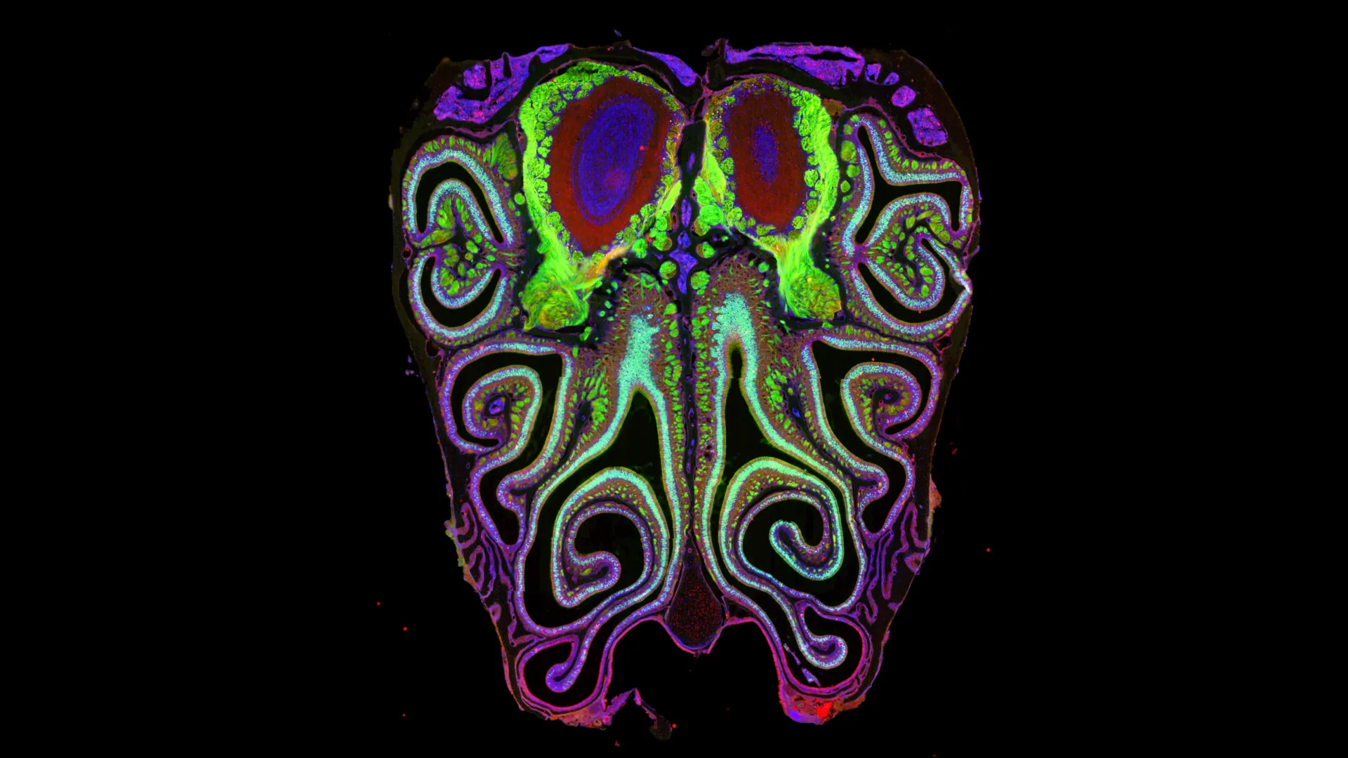
    Smell helps people detect hazards, shapes flavor, and ties closely to memory and emotion, yet its underlying biology has remained harder to map than other senses. New research in mice now outlines a detailed layout of where different smell receptors sit inside the nose.

    The study, published in Cell on April 28, describes the first high-resolution spatial map for more than 1 000 types of olfactory receptors. Researchers say the work challenges the long-held view that these receptor-carrying neurons are arranged largely at random.

    A hidden order in olfaction

    Instead of a scattered pattern, the team found that olfactory sensory neurons form organized horizontal bands across the nasal tissue. These stripes overlap in a consistent way, grouping neurons by the receptor type they express.

    That organization appeared highly reproducible across animals, suggesting it is a stable biological blueprint rather than a quirk of individual development. The researchers also report that the nasal map aligns with corresponding maps in the olfactory bulb, the brain’s first processing hub for smell.

    How the map may form

    To build the atlas, scientists analyzed about 5.5 million neurons collected from more than 300 mice. They combined single-cell sequencing, which identifies receptor identity, with spatial transcriptomics, which preserves information about each cell’s location.

    The team also points to retinoic acid, a molecule known to regulate gene activity, as a key organizer of the pattern. When researchers altered retinoic acid levels, the position of the receptor stripes shifted up or down, indicating a developmental control signal.

    Why it matters for smell loss

    Loss of smell has gained wider attention in recent years, including from post-viral cases, and it can affect safety, nutrition, and mental well-being. Researchers argue that restoring olfaction will require understanding how receptor neurons are positioned and connected to the brain.

    The study’s authors say a clearer map could help guide future efforts to repair damaged smell circuits, including work on cell-based therapies and other interventions. They also note that an important next step is testing whether a similar receptor organization exists in humans.

  • Why Old Buildings Can Feel Unsettling: Researchers Point to Infrasound and Rising Stress Hormones

    Why Old Buildings Can Feel Unsettling: Researchers Point to Infrasound and Rising Stress Hormones

    That uneasy feeling many people report in older buildings may have a measurable physical trigger, according to new research into infrasound, a type of low-frequency vibration that sits below the threshold of human hearing.

    Scientists say these vibrations can be produced by everyday sources such as ventilation systems, traffic, and industrial machinery, and may be especially common in basements where aging pipes and mechanical equipment can generate persistent low-frequency motion.

    In a controlled experiment published in Frontiers in Behavioral Neuroscience, researchers tested whether people could detect infrasound and whether it affected mood. The team focused on 18 Hz, a frequency below 20 Hz that most people cannot consciously hear.

    The study involved 36 participants who sat alone in a room while listening to either calming or unsettling music. For half of the group, hidden subwoofers also generated infrasound during the session.

    Afterward, participants rated how they felt and whether they believed infrasound had been present, and researchers collected saliva samples to measure cortisol, a hormone associated with the body’s stress response. The key question was whether the body would react even when the sound could not be consciously identified.

    Participants exposed to infrasound showed higher salivary cortisol levels and reported feeling more irritable and less engaged, the researchers found. They were also more likely to describe the music as sad, despite being unable to reliably tell whether infrasound had been playing.

    A hidden factor behind haunted vibes

    Researchers argue the results help explain why some spaces feel disturbing without an obvious cause. In an old building, low-frequency vibrations from ventilation or plumbing may subtly influence mood, increasing tension without providing a clear sensory signal to blame.

    The team noted that expectations can shape how people interpret discomfort, such as attributing agitation to paranormal activity after being told a building is haunted. In that context, infrasound offers a non-supernatural mechanism that still produces real, measurable effects.

    What the study does not prove

    The researchers cautioned that the study was relatively small and tested a single frequency over a short exposure, leaving open questions about how different frequencies, combinations, and longer durations might affect people. Real-world infrasound is rarely a clean tone, and its intensity can vary widely by environment.

    They also emphasized that the biological pathway is not yet clear, even though the hormonal and mood shifts were detectable. Future work is expected to examine a broader range of conditions and to track responses during exposure, not only afterward.

    Why long-term exposure matters

    Cortisol plays a normal role in helping the body respond to challenges, but sustained elevation has been linked in broader medical research to health risks, including effects on sleep, mood, and cardiovascular function. The study adds to concerns that chronic, unnoticed low-frequency noise could contribute to ongoing stress in some settings.

    Researchers say clearer evidence could eventually inform building design choices and noise guidelines, particularly around mechanical systems that generate low-frequency vibrations. For now, they suggest that if a room feels inexplicably tense, the cause may be structural and mechanical rather than mysterious.

  • Coffee and the gut-brain axis: New study finds decaf may lift mood and memory too

    Coffee and the gut-brain axis: New study finds decaf may lift mood and memory too

    Scientists in Ireland have reported new evidence that coffee can influence the gut-brain axis, the two-way signaling system linking digestion and brain function. The research suggests both caffeinated and decaffeinated coffee may shape gut microbes and track with changes in mood-related measures.

    The study, led by APC Microbiome Ireland at University College Cork and published in Nature Communications, compared 31 regular coffee drinkers with 31 non-coffee drinkers. Researchers combined diet records, psychological questionnaires, and stool and urine samples to map links between coffee intake, microbiome activity, and wellbeing.

    A two-week coffee break tested

    Regular coffee drinkers were first asked to stop drinking coffee for two weeks, allowing scientists to observe what changed when coffee was removed. During this abstinence period, the team reported shifts in microbe-related metabolites that helped distinguish habitual coffee drinkers from non-drinkers.

    Coffee was then reintroduced in a way that did not tell participants whether they were receiving caffeinated or decaffeinated coffee. According to the findings, both groups showed improvements across several mood measures, including lower reported stress and depressive symptoms.

    Decaf and caffeine showed differences

    One of the more closely watched results was that improvements in learning and memory were seen in the decaffeinated group. The authors say this points to coffee compounds beyond caffeine, such as polyphenols and other bioactives, as possible drivers of certain brain-related effects.

    Meanwhile, the caffeinated group showed changes consistent with caffeine’s known effects, including better alertness and attention, alongside reduced anxiety in the study’s assessments. The paper also reported differences in biological markers tied to inflammation, though it did not position coffee as a treatment.

    What changed in the microbiome?

    The researchers identified specific bacterial signals associated with coffee drinking, including higher levels of Eggertella species and Cryptobacterium curtum in coffee drinkers. They also noted shifts in broader bacterial groupings that have been linked in other research to metabolic and emotional outcomes.

    Experts caution that microbiome studies often show associations rather than direct cause-and-effect, and larger trials are still needed. Still, the work adds to a growing body of evidence that everyday dietary habits, including coffee consumption, may influence both gut activity and mental health.

    In Europe, moderate caffeine intake is generally considered safe for most healthy adults, with guidance often referenced around 400 mg per day from all sources. People who are pregnant, sensitive to caffeine, or managing anxiety, reflux, or sleep issues are typically advised to discuss intake with a clinician.