Category: Psychology

  • MIT researchers map how focused ultrasound could test the brain circuits behind consciousness

    MIT researchers map how focused ultrasound could test the brain circuits behind consciousness

    Scientists at MIT are advancing plans to use transcranial focused ultrasound, a noninvasive technique that can modulate activity in deep brain regions, to study how conscious experience arises. The approach is laid out in a recent roadmap paper that argues the technology can move consciousness research beyond observation and toward direct tests of cause and effect.

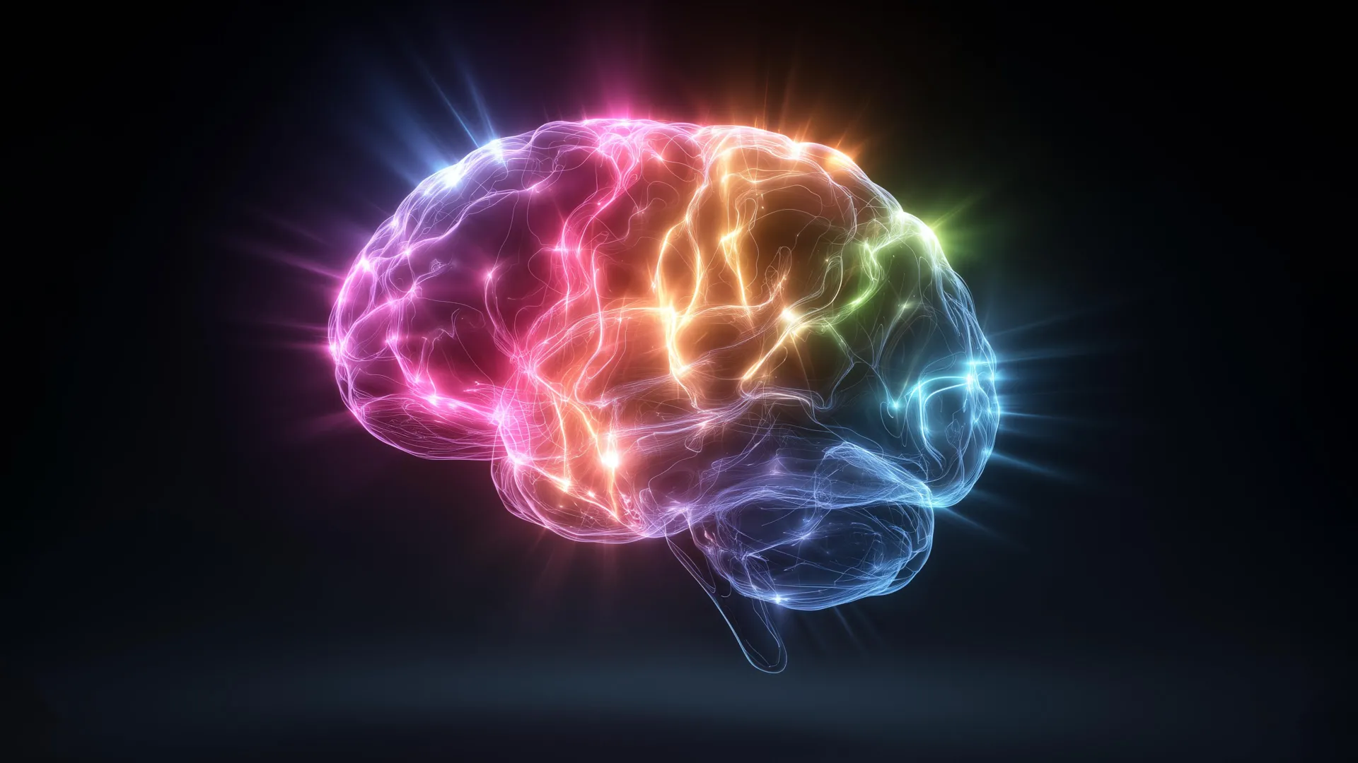
    Consciousness remains a central unsolved problem in neuroscience because most tools either record brain signals or reach only surface areas without surgery. Focused ultrasound can concentrate acoustic energy through the skull onto small targets, potentially enabling precise stimulation of subcortical structures that are difficult to access with other noninvasive methods.

    A tool for cause and effect

    Many studies link conscious perception to patterns seen in EEG or brain imaging, but those signals often show correlation rather than causation. By changing neural activity in a controlled way and tracking what a person reports experiencing, researchers hope to identify which circuits are necessary for awareness and which are downstream side effects.

    MIT researchers Daniel Freeman and Matthias Michel, along with collaborators at the University of Florida and Harvard-affiliated Brigham and Women’s Hospital, argue that focused ultrasound could help narrow the search for the neural substrate of conscious perception. Their paper, published in Neuroscience and Biobehavioral Reviews, describes experimental designs intended for healthy volunteers.

    Testing rival theories of consciousness

    The roadmap highlights how the method could evaluate competing ideas about where consciousness is generated in the brain. Some accounts emphasize higher-level cognitive processes and the prefrontal cortex, while others suggest that specific perceptual regions, posterior networks, or deeper structures may be sufficient to generate conscious experience.

    Because the technology can target areas millimeters across, researchers say it may be possible to compare the effects of stimulating different nodes in these proposed networks. The goal is not only to see brain activity change, but to determine whether those changes reliably alter perception, awareness, or subjective reports.

    From vision to pain and beyond

    Early experiments are expected to start with the visual system, where researchers can tightly control stimuli and measure perception. Similar logic could be applied to pain, a domain where reflexive responses can occur before a person consciously feels discomfort, raising questions about which brain circuits produce the experience itself.

    While focused ultrasound has drawn growing interest for potential therapeutic uses, the authors frame it as a basic-science instrument for probing fundamental mechanisms. They also caution that, as with any emerging method, careful safety standards, calibration, and replication will determine how widely it can be adopted in mainstream neuroscience.

    At MIT, the work is part of a broader push to build a cross-disciplinary community around consciousness research, including regular discussions among neuroscientists and philosophers. For proponents, the appeal is straightforward: a noninvasive way to reach deeper brain targets could provide the most direct experimental leverage the field has had in decades.

  • Parkinson’s trial explores iPSC brain cell implants to restore dopamine and movement

    Parkinson’s trial explores iPSC brain cell implants to restore dopamine and movement

    Doctors at Keck Medicine of USC are taking part in an early-stage clinical trial testing whether implanted stem cell-derived brain cells can help people with Parkinson’s disease regain motor function. The approach aims to replace lost dopamine-producing cells and potentially reduce symptoms driven by dopamine decline.

    Parkinson’s is a progressive neurological condition that affects movement and can also influence mood and cognition. In the United States, more than 1 000 000 people live with the disease, and roughly 90 000 new cases are diagnosed each year, according to recent estimates.

    Aiming to restore dopamine production

    The trial focuses on replenishing dopamine, a key chemical messenger needed for smooth, coordinated movement. As dopamine-producing neurons deteriorate, people may develop tremor, muscle rigidity, slowed movement, and walking and balance difficulties.

    Standard therapies such as levodopa and deep brain stimulation can improve symptoms for many patients, but they do not replace the underlying lost cells. Researchers hope cell replacement could complement existing care by rebuilding the brain’s dopamine-making capacity.

    How the stem cell procedure works

    The study uses induced pluripotent stem cells, or iPSCs, which are adult cells reprogrammed into a flexible state and then guided to become dopamine-producing neurons. Because iPSCs are not embryonic stem cells, the technology is often presented as a less ethically contentious route for creating specialized cells.

    Neurosurgeons implant the cells into the basal ganglia using imaging guidance, aiming for precise placement in a region central to movement control. Participants are monitored closely for 12 to 15 months for safety signals and changes in Parkinson’s symptoms.

    Safety focus and limited enrollment

    Investigators are watching for risks that can follow brain surgery or cell therapies, including infection and abnormal involuntary movements known as dyskinesia. Longer follow-up is expected to continue for up to five years to better understand durability and longer-term safety.

    Keck Medicine is one of three U.S. sites participating in the Phase 1 REPLACE clinical trial, which plans to enroll 12 people with moderate to moderate-severe Parkinson’s disease. The experimental therapy, called RNDP-001, is being developed by Kenai Therapeutics and has received FDA fast-track designation to support an accelerated review pathway if results warrant it.

  • Cambridge study links menopause to grey matter decline, raising new questions about HRT and brain health

    Cambridge study links menopause to grey matter decline, raising new questions about HRT and brain health

    Menopause may be associated with measurable changes in brain structure, alongside higher rates of anxiety, depression and sleep disruption, according to new research led by the University of Cambridge using UK Biobank data.

    In a large sample of women, researchers reported lower grey matter volume in several brain regions after menopause, patterns that were broadly similar whether or not participants had used hormone replacement therapy, commonly known as HRT.

    What the researchers analyzed

    The team examined questionnaire, health and cognitive testing data from nearly 125 000 women in the UK Biobank, a long-running project that links health records with detailed participant assessments.

    They also reviewed brain MRI scans from around 11 000 women, allowing comparisons between those who were pre-menopause, post-menopause without HRT use, and post-menopause with HRT use.

    Mental health and sleep symptoms

    Across the dataset, women who had gone through menopause were more likely to report seeking medical help for anxiety, nervousness or depression, and they were more likely to report persistent sleep problems such as insomnia and fatigue.

    Women who used HRT showed higher levels of anxiety and depression than non-users, but the analysis suggested these differences often existed before menopause, indicating HRT may have been prescribed to people already experiencing symptoms.

    Brain regions tied to memory and emotion

    Imaging results showed reduced grey matter volume after menopause in areas involved in memory and emotional regulation, including the hippocampus, entorhinal cortex and anterior cingulate cortex.

    Because some of these regions are also affected early in Alzheimer’s disease, the findings add to ongoing research into why women are diagnosed with dementia more often than men, though the study does not prove menopause causes dementia.

    On cognitive testing, memory scores were broadly similar across groups, but reaction time tended to be slower after menopause, with evidence that HRT use was associated with a smaller decline in reaction speed.

    The authors emphasized that menopause can be a major health transition and argued for greater attention to mental health support, sleep and lifestyle measures such as exercise and diet, alongside individualized medical advice about HRT.

    The study was published in Psychological Medicine, and the researchers noted that further work is needed to clarify how hormone changes, symptom severity, HRT timing and other health factors interact with brain ageing.

  • Why astringent flavanols in cocoa and berries may help trigger brain activity

    Why astringent flavanols in cocoa and berries may help trigger brain activity

    A dry, puckering sensation from cocoa, some berries and red wine is more than a taste quirk, according to emerging research on flavanols. Scientists are increasingly investigating whether astringency itself can act as a rapid signal to the brain, potentially influencing attention and memory.

    Flavanols are a type of polyphenol long associated in population studies and clinical research with cardiovascular benefits, including improved blood vessel function. They have also been linked to cognitive outcomes, but one persistent challenge is that only a small fraction of consumed flavanols is absorbed into the bloodstream.

    A new focus on taste pathways

    In a recent study in Current Research in Food Science, researchers from Shibaura Institute of Technology in Japan proposed that the sensory experience of astringency may help explain flavanols’ outsized effects. The team hypothesized that stimulation in the mouth could transmit signals through sensory nerves to the central nervous system.

    Working with mice, the researchers administered oral doses of flavanols and compared results with a control group given water. The flavanol groups showed higher activity and exploratory behavior and performed better on learning and memory tasks in the experiments.

    Neurochemistry tied to alertness and stress

    Brain measurements suggested changes in neurotransmitter systems associated with attention and arousal, including dopamine-related activity and the locus coeruleus norepinephrine network. The study also reported shifts in markers linked to sympathetic nervous system activity, which plays a key role in alertness and the body’s stress response.

    The researchers interpreted these patterns as evidence that flavanol-driven astringency may function like a mild physiological challenge, with downstream effects that resemble some aspects of exercise-induced activation. They argue this could help reconcile low bioavailability with observed impacts on brain-related outcomes.

    What it means for everyday diets

    The findings do not mean that any bitter or drying food will reliably boost cognition, and the work is primarily an animal study rather than a clinical trial in humans. Still, it adds momentum to the broader scientific push to understand how sensory cues from food can influence the brain quickly, alongside longer-term effects from digestion and circulation.

    Researchers say the idea could inform future work in sensory nutrition, including how foods might be formulated to balance palatability with measurable physiological responses. For consumers, the most evidence-backed approach remains obtaining flavanol-rich foods as part of an overall healthy diet, rather than treating astringency as a standalone brain hack.

  • Parkinson’s Study Points to SCAN Brain Network as a New Treatment Target, With Early Gains From Non-Invasive Stimulation

    Parkinson’s Study Points to SCAN Brain Network as a New Treatment Target, With Early Gains From Non-Invasive Stimulation

    Researchers say they have identified a specific brain network that may sit at the core of Parkinson’s disease, potentially reshaping how the condition is understood and treated. The findings focus on the somato-cognitive action network, or SCAN, which links planning and thinking with physical movement.

    Parkinson’s is a progressive neurological disorder affecting more than 10 million people worldwide, commonly causing tremor, stiffness, slowed movement, sleep disruption and cognitive changes. Standard therapies such as levodopa can ease symptoms for years, while deep brain stimulation can help selected patients, but neither stops disease progression.

    A network view of Parkinson’s

    The international team, led by China’s Changping Laboratory with collaborators including Washington University School of Medicine in St. Louis, analyzed brain imaging data from more than 800 participants across several centers. The dataset included people with Parkinson’s receiving different treatments, along with healthy volunteers and people with other movement disorders for comparison.

    The researchers report that Parkinson’s is marked by unusually strong connectivity between SCAN and deeper brain structures in the subcortex. Across multiple therapies examined, the study found that treatments tended to work better when they reduced this excessive coupling rather than simply stimulating nearby regions.

    Non-invasive stimulation shows early promise

    Building on that signal, the team tested a personalized, high-precision approach using transcranial magnetic stimulation, a non-invasive technique that delivers magnetic pulses through the scalp. In a small trial, 18 patients who received SCAN-targeted stimulation showed a higher response rate after two weeks than 18 patients who received stimulation near, but not on, the SCAN target.

    In the SCAN-targeted group, 56% met the study’s response threshold, compared with 22% in the comparison group, a roughly 2.5-fold difference. The work suggests that matching stimulation more precisely to an individual’s SCAN anatomy could improve outcomes, though larger and longer studies are needed.

    What comes next for SCAN targeting?

    The authors caution that the study does not prove SCAN changes cause Parkinson’s, and the non-invasive results are early-stage. They also note that more basic research is needed to map how different SCAN subregions relate to specific symptoms such as gait, tremor, mood and cognition.

    Even so, the findings add momentum to efforts to move neuromodulation earlier in the disease course, when symptoms are still developing and disability is lower. The researchers say future trials will explore additional non-invasive approaches, including surface electrode stimulation and low-intensity focused ultrasound, to influence SCAN activity more precisely.

    The study was published in Nature on Feb. 4 and was supported by funding from U.S. National Institutes of Health programs and major Chinese research grants. The authors also disclosed multiple industry relationships and patent interests related to neuromodulation and targeting tools, which were reported as managed under institutional conflict-of-interest policies.

  • Late-life depression could precede Parkinson’s or Lewy body dementia, Danish study suggests

    Late-life depression could precede Parkinson’s or Lewy body dementia, Danish study suggests

    While there is currently no cure for Parkinson’s disease or Lewy body dementia, addressing depression early could improve quality of life and overall care for patients as these diseases develop.

    study published in General Psychiatry provides the most detailed longitudinal evidence to date, demonstrating that depression frequently precedes the diagnosis of PD and LBD and remains elevated for several years thereafter.


    Drawing on comprehensive Danish national health registers, the researchers conducted a retrospective case–control study including 17,711 individuals diagnosed with PD or LBD between 2007 and 2019. Researchers compared these patients with people of similar age and sex who were diagnosed with other long-term conditions, including rheumatoid arthritis, chronic kidney disease, and osteoporosis.


    The results showed a clear pattern: depression occurred more often and earlier in people who went on to develop Parkinson’s disease or Lewy body dementia than in those with other chronic illnesses. In the years leading up to diagnosis, the risk of depression rose steadily, peaking in the three years before diagnosis. Even after diagnosis, patients with Parkinson’s disease or Lewy body dementia continued to experience higher rates of depression than the comparison groups.


    Importantly, this pattern could not be fully explained by the emotional burden of living with a chronic illness. Other long-term diseases that also involve disability did not show the same strong increase in depression risk. This suggests that depression may be linked to early neurodegenerative changes in the brain, rather than being only a psychological reaction to declining health.


    The findings were especially striking for Lewy body dementia, where rates of depression were even higher than in Parkinson’s disease, both before and after diagnosis. Researchers note that differences in disease progression and brain chemistry may help explain this trend.


    “Following a diagnosis of PD or LBD, the persistent higher incidence of depression highlights the need for heightened clinical awareness and systematic screening for depressive symptoms in these patients.” first author Christopher Rohde noted “Thus, our main conclusion—that PD/LBD are associated with a marked excess depression risk preceding and following diagnosis when compared with other chronic conditions—remains valid.”


    The authors emphasize that this does not mean everyone with depression will develop Parkinson’s disease or dementia. Instead, they recommend greater awareness and closer monitoring when depression appears for the first time in older adults.


    While there is currently no cure for Parkinson’s disease or Lewy body dementia, addressing depression early could improve quality of life and overall care for patients as these diseases develop.

  • AI study finds stroke may make the opposite brain hemisphere look younger, offering new clues on recovery

    AI study finds stroke may make the opposite brain hemisphere look younger, offering new clues on recovery

    A new study in The Lancet Digital Health suggests the brain can respond to stroke in a surprising way. Researchers at the USC Mark and Mary Stevens Neuroimaging and Informatics Institute (Stevens INI) found that people with severe physical impairments after a stroke may show signs of a “younger” brain structure in areas that were not damaged. This appears to reflect how the brain adapts and reorganizes itself after injury.

    The research was conducted as part of the Enhancing NeuroImaging Genetics through Meta-Analysis (ENIGMA) Stroke Recovery Working Group. Scientists analyzed brain scans from more than 500 stroke survivors collected across 34 research centers in eight countries. By applying deep learning models trained on tens of thousands of MRI scans, the team estimated the “brain age” of different regions in each hemisphere and examined how stroke affects both structure and recovery.

    “We found that larger strokes accelerate aging in the damaged hemisphere but paradoxically make the opposite side of the brain appear younger,” said Hosung Kim, PhD, associate professor of research neurology at the Keck School of Medicine of USC and co-senior author of the study. “This pattern suggests the brain may be reorganizing itself, essentially rejuvenating undamaged networks to compensate for lost function.”

    AI Reveals Brain Rewiring After Stroke

    To carry out the analysis, researchers used a type of artificial intelligence called a graph convolutional network. This system estimated the biological age of 18 brain regions based on MRI data. They then compared this predicted age with each person’s actual age, a measure known as the brain-predicted age difference (brain-PAD), which serves as an indicator of brain health.

    When these brain age measurements were compared with motor function scores, a clear pattern emerged. Stroke survivors with severe movement impairments, even after more than 6 months of rehabilitation, showed younger-than-expected brain age in regions opposite the site of injury. This effect was especially strong in the frontoparietal network, which plays an important role in movement planning, attention, and coordination.

    “These findings suggest that when stroke damage leads to greater movement loss, undamaged regions on the opposite side of the brain may adapt to help compensate,” Kim explained. “We saw this in the contralesional frontoparietal network, which showed a more ‘youthful’ pattern and is known to support motor planning, attention, and coordination. Rather than indicating full recovery of movement, this pattern may reflect the brain’s attempt to adjust when the damaged motor system can no longer function normally. This gives us a new way to see neuroplasticity that traditional imaging could not capture.”

    Large-Scale Data Reveals Hidden Patterns

    The study relied on ENIGMA, a global collaboration that combines data from more than 50 countries to better understand the brain across different conditions. By standardizing MRI data and clinical information from many research groups, the team created the largest stroke neuroimaging dataset of its kind.

    “By pooling data from hundreds of stroke survivors worldwide and applying cutting-edge AI, we can detect subtle patterns of brain reorganization that would be invisible in smaller studies. These findings of regionally differential brain aging in chronic stroke could eventually guide personalized rehabilitation strategies,” said Arthur W. Toga, PhD, director of the Stevens INI and Provost Professor at USC.

    Toward Personalized Stroke Recovery

    The researchers plan to continue this work by following patients over time, from the early stages after a stroke through long-term recovery. Tracking how brain aging patterns and structural changes evolve could help doctors tailor treatments to each person’s unique recovery process, with the goal of improving outcomes and quality of life.

    Learn more about associations between contralesional neuroplasticity and motor impairment by viewing this video made by the Stevens INI.

    The study, “Deep learning prediction of MRI-based regional brain age reveals contralesional neuroplasticity associated with severe motor impairment in chronic stroke: A worldwide ENIGMA study,” was funded by the National Institutes of Health (NIH) grant R01 NS115845 and supported by international collaborators from institutions including the University of British Columbia, Monash University, Emory University, and the University of Oslo.

  • PET brain scans map ketamine’s rapid antidepressant effect, pointing to a potential biomarker for treatment-resistant depression

    PET brain scans map ketamine’s rapid antidepressant effect, pointing to a potential biomarker for treatment-resistant depression

    Researchers in Japan have reported some of the clearest human evidence yet of how ketamine can relieve symptoms of treatment-resistant depression, using PET brain imaging to track changes in key glutamate receptors. The work adds molecular detail to a treatment already known for acting faster than standard antidepressants.

    Major depressive disorder is a leading cause of disability worldwide, and a substantial share of patients do not improve after trying multiple first-line therapies. For those with treatment-resistant depression, ketamine and the related medicine esketamine have drawn attention because some patients feel relief within hours or days rather than weeks.

    What the PET scans measured

    The study, published in Molecular Psychiatry, used a PET tracer called [11C]K-2 designed to visualize AMPA receptors, proteins that help regulate communication between brain cells. Scientists have long suspected that AMPA receptor activity is central to ketamine’s antidepressant effects, but direct confirmation in living people has been limited.

    The researchers combined data from three clinical trials, comparing 34 patients with treatment-resistant depression against 49 healthy participants. Patients received intravenous ketamine or placebo over a two-week period, with PET scans taken before treatment and after the final infusion.

    Receptor shifts tied to symptom relief

    Before treatment, the PET data suggested patients with treatment-resistant depression had region-specific differences in AMPA receptor availability compared with healthy controls. After ketamine, the brain changes were not uniform, instead appearing as shifts in particular areas involved in mood and reward processing.

    Crucially, the degree and location of AMPA receptor changes tracked with how much a patient’s depressive symptoms improved. The authors highlighted especially notable shifts in regions linked in prior research to depression circuitry, arguing the images provide a direct bridge between earlier animal findings and human clinical response.

    Why it could matter clinically

    If replicated, AMPA receptor PET imaging could become a candidate biomarker to help predict who is most likely to benefit from ketamine, and to guide dosing or treatment strategies. That could be valuable because ketamine response can vary, and clinicians are seeking ways to personalize care while balancing benefit, side effects, and monitoring needs.

    The researchers caution that PET imaging is complex and not widely available, and larger studies would be needed before it could influence routine practice. Still, mapping ketamine’s effects at the receptor level may also support development of new rapid-acting antidepressants that target similar pathways with fewer practical barriers.

  • Study links early depression to brain cell energy changes, hinting at a future blood test

    Study links early depression to brain cell energy changes, hinting at a future blood test

    New research suggests major depressive disorder may be tied to early disruptions in how cells generate and manage energy, a finding that could eventually support earlier and more targeted treatment. Scientists say the results add biological detail to a condition still often diagnosed mainly through symptoms and clinical interviews.

    The study focused on adenosine triphosphate, or ATP, sometimes described as the body’s energy currency because it powers basic cellular work. Researchers examined ATP-related signals in both the brain and blood, aiming to see whether measurable energy patterns track with depression in young adults.

    What the scientists measured

    Teams at the University of Queensland and the University of Minnesota analyzed brain imaging and blood samples from 18 participants aged 18 to 25 diagnosed with major depressive disorder. Their results were compared with samples from people without depression to identify differences linked to the illness.

    According to the researchers, the approach is notable because it looked for matching patterns across the brain and the bloodstream, not just in one system. That raises the possibility that, with more evidence, peripheral markers in blood could one day help flag risk or subtypes of depression earlier.

    An unexpected pattern under stress

    The researchers reported that cells from participants with depression showed higher energy-molecule production while at rest, but had difficulty ramping up energy output when challenged. That stress-response limitation, they argue, could align with common symptoms such as fatigue, slowed thinking, and reduced motivation.

    Scientists involved in the work suggest the pattern may reflect mitochondria that are effectively overcompensating early on, then struggling when demand increases. They caution that the study is small, but say it offers a plausible cellular mechanism worth testing in larger groups.

    What this could mean next

    Major depressive disorder is common and can take years to match with an effective treatment, particularly when fatigue is prominent and persistent. The authors argue that identifying measurable biological signatures could support earlier intervention and more personalized care, rather than trial-and-error alone.

    The research was published in Translational Psychiatry, and the team says follow-up studies are needed to confirm the findings, test whether they predict outcomes, and determine whether they apply across ages and different forms of depression. If replicated, the work could also help frame depression as a whole-body condition with detectable biological changes.

  • Microplastics and the brain: Researchers map possible links to Alzheimer’s and Parkinson’s

    Microplastics and the brain: Researchers map possible links to Alzheimer’s and Parkinson’s

    Microplastics, the tiny plastic fragments found in food, water and household dust, are under growing scrutiny as researchers examine how they may affect the brain. A new scientific review pulls together evidence suggesting these particles could contribute to processes seen in Alzheimer’s and Parkinson’s disease.

    Dementia affects more than 57 million people globally, and experts expect the burden of neurodegenerative disease to rise as populations age. That backdrop is sharpening interest in whether environmental exposures such as microplastics may worsen inflammation or accelerate neurological decline.

    Five mechanisms under scientific review

    The review, published in Molecular and Cellular Biochemistry by an international team including the University of Technology Sydney and Auburn University, outlines several biological routes of potential harm. It focuses on immune activation, oxidative stress, disruption of the blood-brain barrier, mitochondrial dysfunction and direct neuronal injury.

    Researchers argue that if microplastics weaken the blood-brain barrier, the brain may become more vulnerable to inflammatory molecules and immune responses that can damage delicate tissue. In parallel, they describe how oxidative stress could rise if reactive oxygen species increase while antioxidant defenses are depleted.

    Energy disruption and protein buildup concerns

    Another concern highlighted in the paper is mitochondrial interference, which could reduce cellular energy production and strain neurons that rely heavily on steady ATP supply. Over time, energy shortfalls may impair brain function and make nerve cells more susceptible to damage.

    The authors also discuss disease-specific hypotheses, including whether microplastics might promote protein changes associated with Alzheimer’s, such as beta-amyloid and tau accumulation. For Parkinson’s, they note a possible role in α-synuclein aggregation and stress on dopamine-producing neurons.

    What the evidence can and cannot show

    While the review raises plausible pathways, the researchers stress that confirming a direct causal link in humans will require further studies, including exposure measurement and long-term clinical follow-up. Much of the current understanding comes from laboratory and animal research, along with emerging evidence that microplastics can accumulate in organs.

    Even with uncertainties, scientists say practical exposure reduction may be reasonable while research catches up, particularly in everyday food and household contexts. The authors point to reducing reliance on plastic food containers and packaging, limiting plastic-related dust and fibers, and supporting policies that curb plastic pollution at its source.

    Ongoing work at the involved institutions is expected to further test how ingested or inhaled microplastics interact with cells and barriers in the body. Public health experts say clearer answers will depend on standardizing how microplastics are measured and comparing real-world doses across populations.