Category: Psychology

  • Teen Diet and Depression Risk: A Major Review Points to Whole Foods Over Supplements

    Teen Diet and Depression Risk: A Major Review Points to Whole Foods Over Supplements

    A major review led by Swansea University researchers suggests teenagers’ overall diet quality may be linked to mental health, with healthier eating patterns more often associated with fewer depressive symptoms. The findings add to growing interest in nutrition as a modifiable factor that could support adolescent wellbeing.

    Published in the journal Nutrients, the paper assessed evidence from 19 earlier studies examining diet and mental health in adolescents. Across the studies, lower-quality diets were more frequently connected with higher psychological distress, though results varied by design and population.

    Whole-diet patterns stand out

    The review included six randomized controlled trials and 13 prospective cohort studies, allowing the authors to compare different types of evidence. While some studies hinted that specific supplements such as vitamin D might reduce depressive symptoms, the overall picture for individual nutrients was inconsistent.

    By contrast, broader dietary patterns showed clearer and more repeatable signals. The authors argue that focusing on overall balance and quality may be more useful than targeting single nutrients, particularly when translating research into school, family, and public health settings.

    Why adolescence is a key window

    Researchers highlighted adolescence as a critical period for brain development and emotional regulation, when depression symptoms can emerge and become entrenched. Because diet is part of daily life and can be shaped at scale, they see it as a practical area for prevention and early support.

    At the same time, the review notes that diet and mental health do not exist in isolation. Socioeconomic factors and sex differences may influence both what teens eat and how mental health outcomes present, complicating cause-and-effect interpretation.

    What the evidence still misses

    The authors flagged major gaps, including a heavy focus on depression compared with other outcomes such as anxiety, stress, self-esteem, externalizing behavior, and aggression. They also emphasized the need for more standardized methods and improved reporting so results can be compared across studies.

    To strengthen future conclusions, the team proposed a research roadmap that includes exposure-based designs, biological markers, and open science practices. In a statement, corresponding author Hayley Young said, “Overall, our findings suggest that public health and clinical strategies should prioritise whole-diet approaches over isolated supplementation when considering adolescent mental health.”

    The researchers cautioned that more high-quality studies are needed to determine which dietary patterns work best, and for whom. Even so, the review suggests that everyday food choices may play a bigger role in teen mental health than many families and clinicians have assumed.


  • Exercise emerges as a leading option for depression and anxiety, with group programs showing the biggest gains

    Exercise emerges as a leading option for depression and anxiety, with group programs showing the biggest gains

    A major evidence review in the British Journal of Sports Medicine reports that structured exercise can meaningfully reduce symptoms of depression and anxiety, often performing as well as established treatments. The authors assessed a large body of randomized trial data to compare different exercise types, intensities and settings.

    The umbrella review combined results from dozens of prior meta-analyses, covering hundreds of individual trials and tens of thousands of participants across a wide age range. Overall, the synthesis found a medium-sized improvement in depression symptoms and a small-to-medium improvement in anxiety.

    Which workouts seemed most effective

    Aerobic exercise such as running, swimming and dance stood out for depression, particularly when sessions were supervised or done in groups. For anxiety, shorter programs lasting up to about 8 weeks and using lower-intensity activity appeared to deliver the most consistent benefits.

    Researchers also found improvements across resistance training and mind-body approaches such as yoga, tai chi and qigong, as well as mixed programs that combine formats. Effects were observed regardless of sex, suggesting exercise can be broadly useful, even if the best “fit” varies by person.

    Who benefited most in the data

    The strongest reductions were reported among young adults ages 18 to 30 and among women after giving birth, groups that also face elevated risks of mood and anxiety symptoms. The authors note that social and practical factors, including support and accountability, may help explain why group formats performed well.

    While the results were generally comparable to medication or talking therapies, the study does not argue that exercise should replace clinical care for everyone. Instead, it points to exercise as a credible first-line or add-on option, especially where access to therapy or medication is limited or where people prefer non-drug approaches.

    Limits and what comes next

    The authors caution that definitions of intensity, frequency and program length differed across studies, making precise prescriptions harder to standardize. Some age groups and exercise formats were also represented by less pooled data than others.

    Even with those caveats, the review strengthens the case for tailoring exercise to individual needs, including supervision, setting and duration. Clinicians increasingly emphasize that the most effective program is one a person can start safely and sustain, while tracking symptoms and overall wellbeing.

  • Study links sugary drinks to teen anxiety, and researchers say the trend may be harder to reverse than parents think

    Study links sugary drinks to teen anxiety, and researchers say the trend may be harder to reverse than parents think

    A new research review has found a consistent association between high consumption of sugary drinks and increased anxiety symptoms among teenagers. The analysis, led by researchers working with Bournemouth University, was published in the Journal of Human Nutrition and Dietetics.

    The team examined results from multiple earlier studies to see whether patterns held across different adolescent groups. Across the evidence base reviewed, higher intake of sugar-sweetened beverages repeatedly aligned with higher self-reported anxiety symptoms.

    Why drinks are under scrutiny

    Public health efforts have long focused on the physical harms of high-sugar diets, including obesity and type 2 diabetes. The authors argue that mental health outcomes linked to energy-dense, low-nutrient drinks have received comparatively less attention.

    The review covers a wide range of sweetened beverages commonly consumed by teens, including sodas, energy drinks, sweetened juices, and sweetened tea or coffee drinks. Many of these products can deliver large amounts of added sugar quickly, without offering lasting satiety.

    Correlation, not proven causation

    The researchers emphasize that the findings do not prove sugary drinks cause anxiety. Because the review synthesizes mainly observational, survey-based studies, it cannot determine whether sugary beverages lead to anxiety or whether anxious teens are more likely to consume them.

    They also note that shared factors could influence both outcomes, such as sleep problems, family stress, or broader lifestyle patterns. The authors say future research designed to test cause-and-effect is needed before drawing firm conclusions.

    What this could mean for families

    Even without proof of causality, the authors say the repeated link is concerning at a time when youth mental health problems are widely reported to be rising. They suggest sugary drink intake is a modifiable habit that could be considered alongside other daily factors like sleep, physical activity, and overall diet quality.

    The review’s lead author, Dr. Karim Khaled, previously completed doctoral work at Bournemouth University and now works at Lebanese American University in Beirut. Co-author Dr. Chloe Casey said mental health effects of diet deserve closer consideration in adolescent nutrition strategies.

  • Brain development may extend into your early 30s, reshaping the age 25 myth

    Brain development may extend into your early 30s, reshaping the age 25 myth

    The popular claim that the brain finishes maturing at 25 has become a shorthand explanation on social media for impulsive decisions and uneven judgment in young adulthood. Neuroscientists say the idea is rooted in older imaging work, but it oversimplifies a longer and more gradual process.

    Earlier longitudinal MRI studies tracked changes in grey matter across childhood and adolescence, including synaptic pruning that strengthens frequently used circuits and trims others. Because many of those datasets ended in the early 20s, age 25 emerged as a convenient estimate rather than a hard biological cutoff.

    New scans map longer maturation

    More recent research has shifted from single brain regions to how whole networks communicate via white matter, the fiber pathways that connect distant areas. In a large analysis spanning infancy to old age, researchers identified a prolonged phase of network reorganization that can stretch into the early 30s.

    One focus is how the brain balances segregation and integration as it matures. Segregation builds specialized clusters for different functions, while integration strengthens long-range connections that help coordinate complex tasks like planning and self-control.

    What changes after the early 30s

    Data suggest a turning point around the early 30s, when some developmental trends level off and the brain shifts toward maintaining well-used pathways. Rather than continuous expansion, the emphasis becomes efficiency and stability in the connections a person relies on most.

    Experts caution that labeling adults as unfinished can be misleading, because brain development is not a single finish line and varies across individuals. Maturation also depends on experience, education, sleep, stress, and health, all of which can shape brain organization over time.

    Why the finding matters

    The updated picture supports a more nuanced view of neuroplasticity, the brain’s ability to adapt across the lifespan. While younger brains often show faster structural change, learning and training remain possible well beyond the 20s, and there is no sudden switch that flips at 25.

    Researchers say the takeaway is not that people in their late 20s are incapable of adult judgment, but that key neural systems may still be refining how efficiently they communicate. That may help explain why many people report steadier decision-making and emotional regulation as they move through their 30s.

  • Parkinson’s trial tests dopamine stem cell implants at USC, offering a closer look at brain repair

    Parkinson’s trial tests dopamine stem cell implants at USC, offering a closer look at brain repair

    Doctors at Keck Medicine of USC have begun implanting dopamine-producing stem cells into the brains of people with Parkinson’s disease, as part of an early-stage clinical trial designed to assess safety and feasibility. The approach aims to replace nerve cells lost to the disorder and restore dopamine production at its source.

    Parkinson’s is a progressive neurological condition best known for tremor, stiffness and slowed movement, but it can also affect sleep, mood and cognition. In the United States, more than 1 000 000 people are living with Parkinson’s, and roughly 90 000 new cases are diagnosed each year, according to public health estimates.

    Why dopamine loss matters

    The disease is closely tied to the gradual death of dopamine-producing neurons, which disrupts signals that help coordinate movement. Standard treatments, including levodopa and other medications, can reduce symptoms, but they do not reliably stop the underlying neurodegeneration.

    The new strategy attempts to address that core deficit by placing replacement cells directly into brain circuits involved in motor control. Researchers say that if transplanted cells survive and produce dopamine in a regulated way, patients could see more stable symptom control than medication alone can provide.

    How the stem cell procedure works

    The trial uses induced pluripotent stem cells, or iPSCs, which are created by reprogramming adult cells back into a flexible state that can be guided to become specific cell types. In this study, the cells are engineered to mature into dopamine-producing neurons intended to integrate into the brain.

    During surgery, clinicians create a small opening in the skull and use imaging guidance to deliver the cells into the basal ganglia, a region central to movement regulation. Participants are monitored closely after implantation for complications such as infection and abnormal involuntary movements, known as dyskinesia.

    What researchers will watch next

    The multi-site U.S. study plans to enroll 12 people with moderate to moderate-severe Parkinson’s disease, with follow-up in the first 12 to 15 months focused heavily on safety signals. Researchers also plan longer observation, up to five years, to evaluate durability and potential longer-term risks.

    The investigational therapy, called RNDP-001, is produced by Kenai Therapeutics, and the FDA has granted the study fast-track designation, a process meant to speed development for serious conditions with unmet need. Even with that designation, researchers caution that larger trials would be required to determine whether the implants provide consistent, meaningful clinical benefit.

    Stem cell and cell-replacement approaches are an active area of Parkinson’s research worldwide, reflecting a broader push to move beyond symptom relief toward disease-modifying strategies. For now, the USC-led effort adds closely watched clinical data on whether implanted dopamine neurons can be delivered safely and function as intended in patients.

  • New Alzheimer’s study points to subtle brain blood flow changes as an early warning sign

    New Alzheimer’s study points to subtle brain blood flow changes as an early warning sign

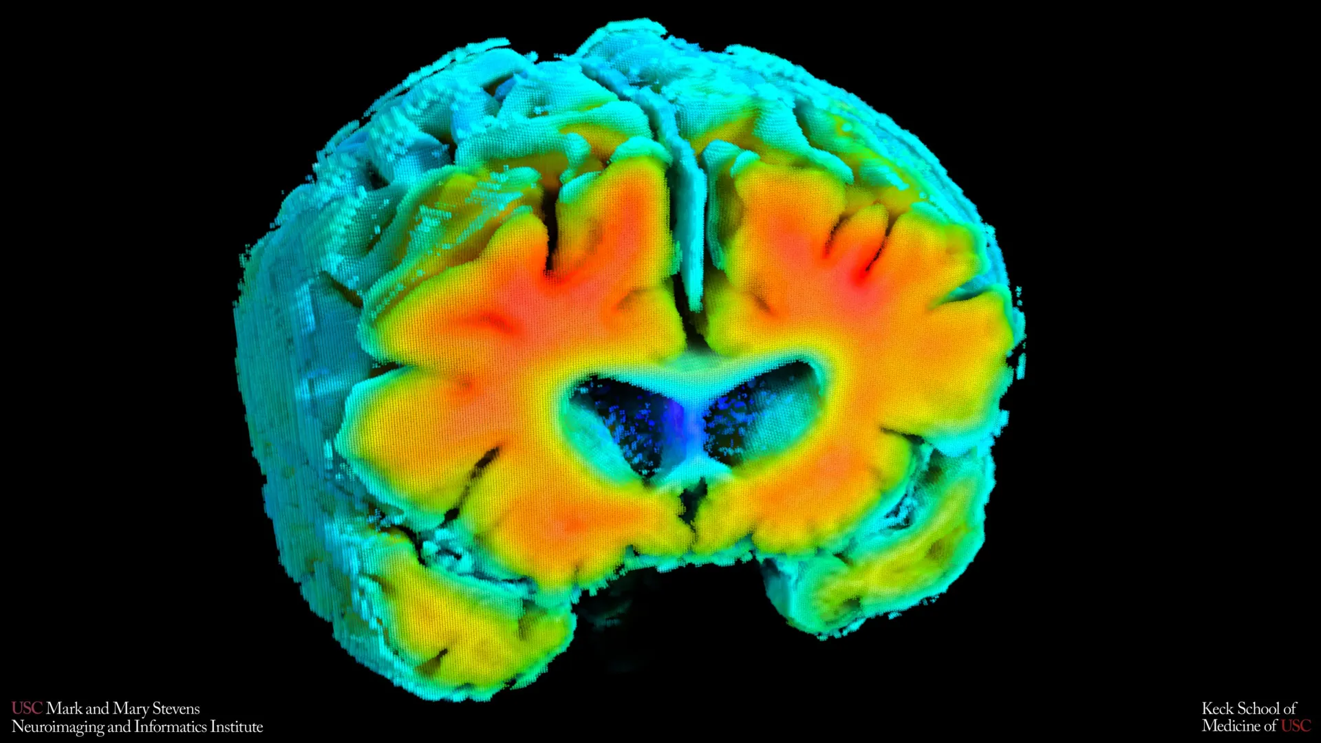
    Small shifts in how blood moves through the brain and how brain cells receive oxygen may be closely connected to the risk of Alzheimer’s disease. That is the conclusion of new research from the Mark and Mary Stevens Neuroimaging and Informatics Institute (Stevens INI) at the Keck School of Medicine of USC.

    The study, published in Alzheimer’s and Dementia: The Journal of the Alzheimer’s Association, examined older adults both with and without cognitive impairment. Researchers found that simple, noninvasive measures of brain blood flow and oxygen levels were linked to well known signs of Alzheimer’s, including amyloid plaque buildup and shrinkage of the hippocampus, the part of the brain that plays a central role in memory. The results suggest that the health of the brain’s blood vessels may influence the disease process early on and could help flag people at risk before noticeable symptoms develop.

    “Amyloid and tau are often considered the primary players in Alzheimer’s disease, but blood flow and oxygen delivery are also critical,” said Amaryllis A. Tsiknia, lead author of the study and USC PhD candidate. “Our results show that when the brain’s vascular system functions more like it does in healthy aging, we also see brain features that are linked to better cognitive health.”

    Noninvasive Tools to Measure Brain Circulation

    To study these changes, the team relied on two painless techniques that can be used while a person rests quietly. Transcranial Doppler ultrasound tracks how quickly blood travels through the brain’s major arteries. Near infrared spectroscopy evaluates how effectively oxygen reaches brain tissue near the surface of the cortex.

    Researchers then applied advanced mathematical modeling to combine these readings into overall indicators of cerebrovascular function. These indicators reflect how well the brain adjusts blood flow and oxygen delivery in response to natural fluctuations in blood pressure and carbon dioxide.

    Vascular Health Linked to Amyloid and Memory Centers

    Participants whose vascular indicators more closely resembled those of cognitively healthy adults tended to have lower amyloid levels and a larger hippocampus. Both features are associated with reduced Alzheimer’s risk.

    “These vascular measures are capturing something meaningful about brain health,” said Meredith N. Braskie, PhD, senior author of the study and assistant professor of neurology at the Keck School of Medicine. “They appear to align with what we see on MRI and PET scans that are commonly used to study Alzheimer’s disease, providing important information about how vascular health and standard brain measures of Alzheimer’s disease risk may be related.”

    The researchers also observed that people diagnosed with mild cognitive impairment or dementia showed weaker vascular function compared to cognitively normal participants. This finding supports the view that declining blood vessel health in the brain is part of the broader Alzheimer’s disease continuum.

    “These findings add to growing evidence that Alzheimer’s involves meaningful vascular contributions in addition to classic neurodegenerative changes,” said Arthur W. Toga, PhD, director of the Stevens INI. “Understanding how blood flow and oxygen regulation interact with amyloid and brain structure opens new doors for early detection and potentially prevention.”

    Potential for Earlier and Broader Screening

    Compared with MRI and PET imaging, these methods are less costly and easier to perform. They do not involve injections, radiation exposure, or demanding tasks for patients. That simplicity could make them useful for large scale screening or for individuals who are unable to undergo more intensive brain imaging.

    The authors caution that the findings represent a single snapshot in time and do not establish cause and effect. Ongoing long term studies are tracking participants to see whether shifts in these vascular measures can predict future cognitive decline or response to treatment.

    “If we can track these signals over time, we may be able to identify people at higher risk earlier and test whether improving vascular health can slow or reduce Alzheimer’s-related brain changes,” Tsiknia said.

    About the Study

    In addition to Tsiknia and Braskie, the study’s other authors are Peter S. Conti, Rebecca J. Lepping, Brendan J. Kelley, Rong Zhang, Sandra A. Billinger, Helena C. Chui and Vasilis Z. Marmarelis.

    This work was supported by the Office of The Director, National Institutes of Health, under Award Number S10OD032285, and by the National Institute on Aging [R01AG058162].

  • UCLA study suggests accelerated TMS may ease treatment-resistant depression in five days, but follow-up timing matters

    UCLA study suggests accelerated TMS may ease treatment-resistant depression in five days, but follow-up timing matters

    Researchers at UCLA Health say an accelerated form of transcranial magnetic stimulation, or TMS, may deliver comparable symptom relief for some people with treatment-resistant depression in just five days. The approach aims to reduce the practical burden of the standard schedule, which typically requires daily weekday visits over six to eight weeks.

    TMS is a noninvasive brain stimulation therapy that uses magnetic pulses to target regions involved in mood regulation. It is commonly offered when patients do not improve after multiple antidepressant trials, and it has become a widely used option in outpatient psychiatric care.

    A five-by-five TMS schedule

    In the study, clinicians compared a compressed protocol known as 5×5, meaning five sessions per day for five consecutive days, with a conventional six-week course. The analysis included 175 patients, with 40 receiving the accelerated format and 135 receiving standard treatment.

    Both groups saw significant reductions in depression symptoms, and overall outcomes were not meaningfully different between schedules. The findings were published in the Journal of Affective Disorders, and the authors noted the results could expand access for patients who struggle to attend weeks of appointments.

    Why follow-up may change results

    One key observation was that some patients in the accelerated group did not appear to improve immediately after the five-day course ended. When assessed again two to four weeks later, those individuals showed substantial improvement, with depression scores falling by an average of 36%.

    That pattern suggests end-of-week assessments may underestimate the benefit of accelerated TMS for certain patients. The researchers said timing could be important when clinicians and patients decide whether a short course is working.

    What the study did not prove

    The researchers cautioned that the comparison was not a randomized clinical trial, meaning patients were not randomly assigned to the accelerated or standard schedule. They also reported that the traditional course performed better on some longer-term measures, underscoring the need for larger controlled trials.

    Even so, the team argues that a shorter TMS pathway could help more eligible patients start and complete care, especially those facing transportation, work, or caregiving barriers. Further research is expected to refine who benefits most, and whether booster sessions after a short course improve durability.

  • NYU Study Links Aging Anxiety to Faster Biological Aging in Women, with Health Worries Playing the Biggest Role

    NYU Study Links Aging Anxiety to Faster Biological Aging in Women, with Health Worries Playing the Biggest Role

    Worrying about getting older, particularly fears of future health decline, may be tied to faster biological aging in women, according to new research from NYU School of Global Public Health. The study connects aging anxiety with changes in epigenetic markers measured in blood.

    Researchers analyzed data from 726 women in the Midlife in the United States study, combining survey responses with laboratory measures. They focused on whether subjective anxiety about aging aligned with objective indicators of cellular aging.

    How biological aging was measured

    The team used two widely cited epigenetic clocks to assess aging biology, including DunedinPACE, which estimates the pace of aging, and GrimAge2, which is designed to capture accumulated biological risk. Higher anxiety scores were associated with faster aging on DunedinPACE.

    The clearest link involved worries about age-related health problems rather than concerns about appearance or fertility. Researchers suggested health fears may be more persistent over time, potentially making them more relevant to longer-term stress biology.

    Why the link may matter

    Accelerated epigenetic aging has been associated in prior research with higher risk of age-related disease and earlier functional decline, though it is not a diagnosis on its own. The NYU findings add to evidence that psychological stressors can correlate with measurable biological changes.

    The authors emphasized that mental and physical health are often treated separately despite frequent overlap. They argued that addressing aging anxiety could be a meaningful, potentially modifiable factor in supporting healthier aging.

    Limits and what comes next

    The study captured a single point in time, so it cannot prove that anxiety causes faster aging. When researchers adjusted for health behaviors that can accompany anxiety, such as smoking and alcohol use, the statistical link weakened and was no longer significant.

    The team called for further research to clarify cause and effect, track changes over time, and identify which interventions might reduce harmful stress while supporting healthier coping. They also pointed to broader social pressures and caregiving burdens that can intensify midlife worries for women.

  • AI-built molecular atlas maps Alzheimer’s brain beyond amyloid plaques, pointing to overlooked metabolic shifts

    AI-built molecular atlas maps Alzheimer’s brain beyond amyloid plaques, pointing to overlooked metabolic shifts

    Researchers at Rice University have created a label-free molecular atlas of the Alzheimer’s brain in an animal model, using laser-based imaging paired with artificial intelligence. The work aims to clarify how the disease emerges and spreads beyond what standard pathology typically captures.

    The study used hyperspectral Raman imaging, an advanced form of Raman spectroscopy that reads chemical fingerprints in tissue without dyes or fluorescent tags. By scanning brain slices at high resolution, the team generated a detailed chemical map designed to reflect the brain’s native state.

    What the imaging revealed

    Analysis indicated that Alzheimer’s-linked chemical changes were not limited to amyloid plaques. Instead, the alterations appeared across multiple brain regions, with uneven patterns that could help explain why symptoms develop gradually and differ between individuals.

    To handle the large dataset, the researchers applied both unsupervised and supervised machine learning methods. Unsupervised tools grouped tissue by molecular similarity, while supervised models helped distinguish Alzheimer’s-affected samples from controls across different regions.

    Metabolic signals in key regions

    Beyond protein-related pathology, the maps pointed to broader metabolic differences, including shifts in cholesterol and glycogen signals. The strongest contrasts were reported in brain regions central to memory and cognition, including the hippocampus and cortex.

    The authors argue that these molecular patterns support a wider view of Alzheimer’s as a disorder involving disrupted brain structure and energy balance, not only plaque formation. They say a whole-brain, label-free approach could help surface changes that targeted assays might miss.

    While the findings are based on an animal model and would need validation in human tissue, the researchers suggest the approach could eventually inform earlier detection strategies and more region-specific treatment research. The study was published in ACS Applied Materials and Interfaces with support from U.S. federal research funders.

  • Study identifies DeltaFosB in the hippocampus as a key driver of cocaine relapse, opening a path to targeted treatments

    Study identifies DeltaFosB in the hippocampus as a key driver of cocaine relapse, opening a path to targeted treatments

    Scientists at Michigan State University have pinpointed a brain protein that appears to be essential for the circuit changes that fuel cocaine relapse, offering a clearer biological explanation for why cravings can persist long after use stops. The findings, published in Science Advances and supported by the US National Institutes of Health, focus on a molecule called DeltaFosB.

    The research highlights the hippocampus, a region central to memory and learning, and its interaction with reward pathways involved in drug seeking. By linking relapse risk to durable changes in these circuits, the study adds weight to the view that cocaine addiction is driven by brain biology rather than willpower alone.

    How cocaine rewires memory circuits

    Unlike opioids, stopping cocaine does not typically cause severe physical withdrawal, yet relapse remains common, and no FDA-approved medication is specifically indicated for cocaine use disorder. Cocaine’s surge of dopamine reinforces drug-taking, while memory-linked cues can later reignite the urge to use.

    Using mouse models and a specialized CRISPR-based approach, the team found that DeltaFosB acts like a genetic switch in a pathway connecting reward centers and the hippocampus. With repeated cocaine exposure, DeltaFosB accumulates and changes how neurons respond, increasing the drive to seek the drug.

    Genes that intensify cocaine seeking

    The researchers also identified genes influenced by DeltaFosB after longer-term cocaine exposure, including calreticulin, which helps regulate how neurons communicate. In experiments, higher calreticulin activity appeared to boost signaling in pathways that promote continued cocaine seeking.

    Crucially, the study suggests DeltaFosB is not merely associated with these adaptations but required for them to fully develop. When the protein’s role was disrupted, cocaine did not produce the same patterns of brain activity changes linked to persistent drug seeking.

    What this could mean for treatment

    Because many of the implicated genes and circuits are conserved across mammals, the authors say the findings could help guide human research, though direct clinical implications remain years away. The group is now collaborating with the University of Texas Medical Branch to develop compounds aimed at altering how DeltaFosB binds to DNA.

    Future work will also explore how hormones may shape these circuits and whether addiction-related brain adaptations differ between males and females. Such insights could eventually support more personalized approaches to treating cocaine use disorder.