Blog

  • A lasting brain switch in addiction and stress: Why ΔFosB is drawing new attention in mental health research

    A lasting brain switch in addiction and stress: Why ΔFosB is drawing new attention in mental health research

    Advances in molecular psychiatry are sharpening scientists’ view of how stress and drugs can leave long-lasting marks on the brain. A recent interview published by Genomic Press in the journal Brain Medicine highlights decades of work that helped connect fleeting experiences to persistent changes in behavior.

    In the discussion, neuroscientist Eric J. Nestler, dean of the Icahn School of Medicine at Mount Sinai, traces how early training in brain chemistry led him to focus on the biology behind addiction, depression and resilience. He describes a field that has moved from broad theories toward specific molecules, cell types and circuits that can be measured.

    ΔFosB and long-term brain change

    One of the best-known findings from this line of research centers on ΔFosB, a transcription factor that can build up in reward-related brain circuits after repeated drug exposure and prolonged stress. Unlike many proteins that degrade quickly, ΔFosB can persist for weeks, helping to explain how short periods of exposure may trigger longer-lasting shifts in gene activity.

    Researchers have linked this durability to changes in motivation and reward processing that can raise vulnerability to addiction. The idea is not that one factor explains complex disorders, but that stable molecular signals like ΔFosB can act as a biological bridge between experience and enduring neural adaptation.

    From epigenetics to single-cell tools

    Nestler also points to a major methodological shift in the field, from studying signaling pathways to mapping gene regulation through epigenetic mechanisms such as chromatin modifications. Those approaches have been accelerated by tools that can parse differences across brain regions and, increasingly, across individual neuron types.

    Single-cell methods are now enabling researchers to look for patterns that may be missed when tissue is analyzed in bulk. That trajectory is feeding interest in whether future treatments could be tailored more precisely to particular circuits or cell populations involved in mood and substance-use disorders.

    Why resilience is becoming central

    A notable theme in the interview is a push to study resilience, not only pathology. Experiments in animals have identified molecular and circuit signatures associated with maintaining normal behavior despite stress, raising the possibility of therapies designed to strengthen protective mechanisms.

    Some resilience-oriented strategies are already being tested clinically for depression, reflecting a broader shift toward interventions that aim to improve adaptive capacity as well as relieve symptoms. The interview argues that focusing on what helps certain individuals recover could open complementary pathways for drug development.

    Nestler also underscores the importance of linking animal findings with human evidence, including results from postmortem brain studies in people affected by addiction and stress-related conditions. He warns that politicizing science could slow progress, stressing that medical research should remain independent and broadly beneficial.

  • Astrocytes move into the spotlight: New Nature study links overlooked brain cells to fear memories and PTSD pathways

    Astrocytes move into the spotlight: New Nature study links overlooked brain cells to fear memories and PTSD pathways

    Brain research is increasingly challenging the long-held idea that neurons alone drive fear and trauma responses. A new study in Nature points to astrocytes, star-shaped support cells, as active players in how fear memories are formed, recalled and reduced.

    Astrocytes are widely distributed throughout the brain and have traditionally been seen as caretakers that keep neural circuits stable. The new work suggests they can also shape signaling in the amygdala, a central hub for processing threat and generating fear-related learning.

    What the researchers observed

    Using a mouse model of fear learning, scientists tracked astrocyte activity in real time with fluorescent sensors. Astrocyte signaling rose during fear conditioning and again during memory recall, then declined as fear responses weakened through extinction training.

    The team also manipulated how astrocytes communicate with nearby neurons. Enhancing astrocyte-to-neuron signaling strengthened fear expression, while dampening those signals reduced fear responses, indicating astrocytes can tune the intensity of fear memories.

    How it changes the fear circuit

    When astrocyte activity was disrupted, neurons in the amygdala had difficulty forming the typical activity patterns associated with fear. That interference also appeared to affect how defensive-response information is routed to other brain regions involved in choosing and executing behavior.

    Researchers reported effects beyond the amygdala, including changes in fear-related signaling reaching the prefrontal cortex, an area tied to decision-making and regulation of emotional responses. The results suggest astrocytes may influence how the brain decides whether a threat response is appropriate.

    Why it matters for PTSD

    PTSD and several anxiety disorders are marked by persistent, hard-to-extinguish fear memories and heightened reactions to cues that are no longer dangerous. If astrocytes help govern both the expression and the fading of fear, they could become a complementary target alongside neuron-focused approaches.

    The researchers caution that translating mouse findings to human treatments takes time, but the study reframes fear circuitry as a partnership between neurons and glia. Next steps include mapping astrocyte roles across the wider threat network, including regions that coordinate freezing and flight responses.

  • Warm hugs and the brain: New thermoception research reveals how temperature shapes body awareness and mood

    Warm hugs and the brain: New thermoception research reveals how temperature shapes body awareness and mood

    New neuroscience research is sharpening the picture of why a warm hug can feel uniquely calming, suggesting that temperature signals from the skin help the brain maintain a stable sense of the body. A recent review argues that thermoception, the ability to sense warmth and cold, plays a larger role in emotion and self-awareness than previously assumed.

    Published in Trends in Cognitive Sciences, the review brings together evidence from psychology, neuroscience and clinical studies to show that temperature is not just about comfort or survival. Instead, thermal cues appear to influence how strongly people experience their body as their own, a process often described as body ownership.

    Temperature as a body-brain signal

    Researchers highlight that thermoception works alongside touch and internal bodily signals to shape moment-to-moment awareness of the self. Warmth in particular is framed as a biologically meaningful cue of safety and care, learned early in life and reinforced through social contact.

    Laboratory work has linked warm, gentle contact to neural pathways that feed into brain regions involved in interoception, including the insular cortex. These circuits help integrate what the body feels from the outside with internal state, supporting emotional regulation during close social interactions.

    Links to mental health conditions

    The review points to clinical observations in which disrupted body awareness is common, including depression, anxiety, trauma-related disorders and eating disorders. In these settings, people may describe feeling detached from their body or less certain about bodily sensations.

    Studies in conditions such as stroke, anorexia nervosa and body integrity dysphoria suggest that altered thermal perception can occur alongside disturbances in body ownership. The authors argue this overlap makes temperature-sensing a promising, if underused, lens for understanding symptoms that involve disconnection from the body.

    From therapy to prosthetics

    Beyond explaining everyday comfort, the authors suggest thermoception research could inform sensory-based approaches in rehabilitation and mental health care. Better mapping of skin-to-brain temperature pathways may help clinicians identify vulnerabilities and tailor interventions that work through controlled sensory input.

    Engineers could also apply these insights to prosthetics, where adding realistic thermal feedback may improve how naturally an artificial limb is experienced. The review further notes that more frequent exposure to extreme heat and cold could affect mood and stress, making temperature an emerging topic in public health research.

    In practical terms, the science helps explain why warm social touch can be grounding: thermal and tactile signals arrive together, reinforcing the brain’s model of the body in a context associated with safety. That combination may be one reason a brief, warm hug can feel like both physical comfort and emotional reassurance.

  • UCSF study flags FTL1 protein as a driver of brain aging, and a potential new target to restore memory

    UCSF study flags FTL1 protein as a driver of brain aging, and a potential new target to restore memory

    Aging can take a heavy toll on the hippocampus, the brain region central to learning and memory. Researchers at the University of California, San Francisco report they have identified a protein that may play an outsized role in that decline.

    In a study published in Nature Aging, the team points to FTL1, a ferritin-related protein involved in iron handling, as a key molecular change seen in older mouse hippocampus. The researchers say shifting FTL1 levels altered memory performance and the strength of neural connections in ways that tracked with age.

    A standout signal in aging brains

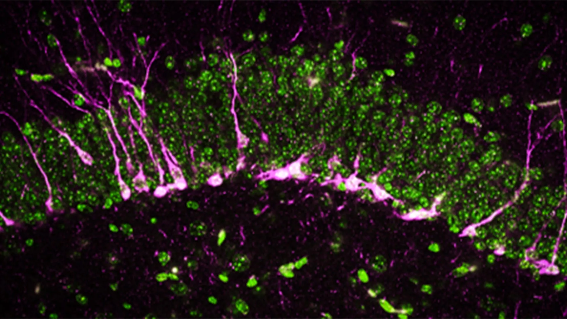
    To pinpoint what changes over time, scientists compared gene and protein patterns in the hippocampus of young and older mice. Among many measurements, FTL1 emerged as the most consistent difference between age groups.

    Older mice had higher FTL1 levels alongside fewer synaptic connections and worse results on cognitive tasks. The authors report that the pattern suggested more than a passive marker of aging, raising the possibility that FTL1 helps drive the process.

    What happened when FTL1 was altered

    When researchers increased FTL1 in young mice, the animals developed brain and behavioral changes resembling those seen in older mice. The hippocampus showed reduced connectivity, and performance on memory-related testing declined.

    Cell experiments offered a potential explanation: neurons pushed to make more FTL1 formed simpler structures, with fewer branching extensions needed for complex signaling. That shift, the team argues, could help explain how elevated FTL1 weakens hippocampal circuitry.

    Can memory decline be reversed?

    In older mice, lowering FTL1 was linked to improved synaptic connections and better memory test performance. Senior author Saul Villeda said, “It is truly a reversal of impairments,” emphasizing that the effect went beyond delaying decline.

    The group also reported a metabolic component, with higher FTL1 associated with slower energy use in hippocampal cells. In lab settings, boosting cellular metabolism with an experimental compound reduced the harmful effects tied to elevated FTL1, pointing to possible therapeutic angles.

    Experts caution that mouse findings do not automatically translate to human brain aging, and any treatment approach would require extensive safety and efficacy testing. Still, the UCSF team argues that targeting FTL1 or related metabolic pathways could eventually open a new route for interventions aimed at age-related cognitive decline.

  • Japanese researchers grow thalamus-cortex brain circuits in lab assembloids, opening a new window on neural development

    Japanese researchers grow thalamus-cortex brain circuits in lab assembloids, opening a new window on neural development

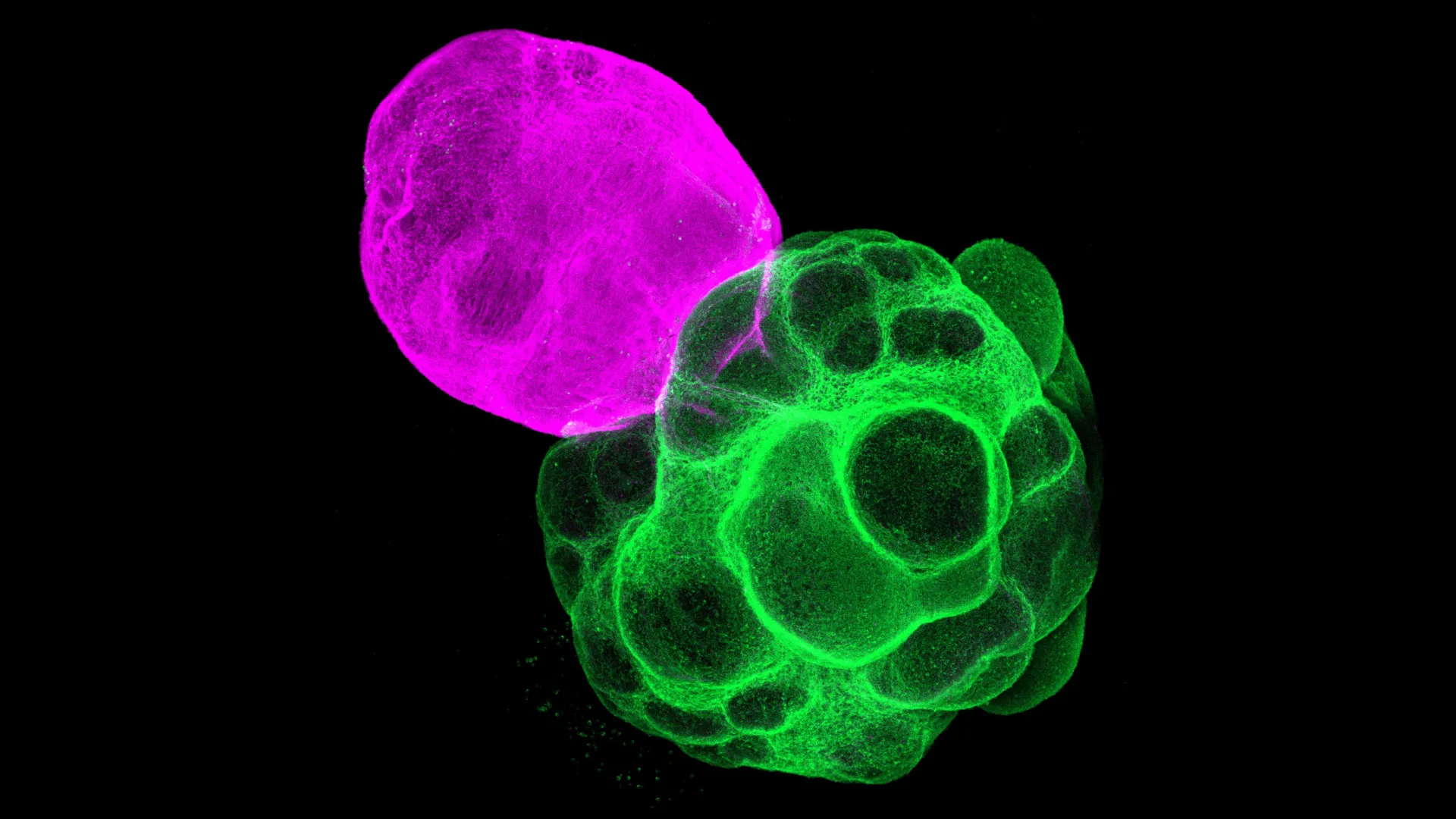
    Researchers in Japan have recreated key human brain circuits in the lab by fusing miniature models of the thalamus and cerebral cortex into what are known as assembloids. Built from human induced pluripotent stem cells, the multi-region tissue is designed to mimic how developing brain areas connect and coordinate activity.

    The work, published in the Proceedings of the National Academy of Sciences, focuses on the thalamus-cortex pathway that helps organize sensory processing and higher cognition. Because direct studies of early human brain wiring are limited by ethical and technical constraints, organoid-based models have become an important alternative.

    Why thalamus-cortex wiring matters

    The cerebral cortex relies on precisely timed communication among different neuron types and between distant brain regions. Disruptions in these networks are linked to neurodevelopmental conditions, including autism spectrum disorder, making circuit formation a high-priority target for basic and translational research.

    Animal studies have long suggested the thalamus helps shape cortical development, but confirming the details in human tissue has been difficult. The new assembloid model aims to capture those cross-region interactions more realistically than single-region organoids.

    How the assembloids were built

    The team first grew separate thalamic and cortical organoids from iPS cells and then fused them to allow fibers to extend between regions. Over time, axons from each side grew toward the other and formed synapses, resembling the bidirectional connectivity seen in the developing brain.

    When researchers compared cortical tissue grown alone with cortical tissue connected to the thalamus, the connected cortex showed gene activity consistent with greater maturation. The results support the idea that thalamic input can accelerate aspects of cortical development.

    Signals that synchronize specific neurons

    To probe function, the scientists tracked how activity traveled through the fused tissue and observed wave-like patterns moving from the thalamus into the cortex. That flow was associated with synchronized activity across parts of the cortical network.

    The effect differed across major cortical excitatory neuron classes, with synchronized patterns seen in pyramidal tract and corticothalamic neurons that communicate with the thalamus. Intratelencephalic neurons, which do not project to the thalamus, showed less synchrony, suggesting thalamic input selectively reinforces certain circuit types.

    Researchers say the platform could help clarify how human circuits assemble at the level of cell types and connections, and provide a testbed for studying disease mechanisms. In the longer term, thalamus-cortex assembloids may also support screening strategies aimed at restoring or stabilizing abnormal network activity.

  • Researchers map a brain pathway that may signal fullness: Astrocytes emerge as a new appetite control target

    Researchers map a brain pathway that may signal fullness: Astrocytes emerge as a new appetite control target

    Scientists have identified a previously underappreciated brain signaling pathway that helps the body recognize when it is time to stop eating, shifting attention from neurons alone to a broader cellular network. The work, published April 6, 2026 in Proceedings of the National Academy of Sciences, focuses on how the hypothalamus processes post-meal fuel signals.

    The study centers on astrocytes, abundant brain cells long viewed mainly as support for neurons, and suggests they can actively shape appetite control. Researchers say this mechanism could eventually inform new strategies for obesity and eating-disorder treatments, though the findings are based on animal experiments.

    How glucose signals reach the brain

    After a meal, glucose levels rise and are sensed in part by tanycytes, specialized cells that line fluid-filled spaces in the brain. In the experiments, tanycytes responded to glucose by producing lactate, a metabolic byproduct that can function as a signaling molecule in the surrounding tissue.

    For years, lactate was often discussed as a signal that could act directly on appetite-regulating neurons. This research argues the message commonly takes an additional step, with astrocytes serving as a crucial intermediary before neurons that promote satiety are engaged.

    Astrocytes as appetite messengers

    The team found that astrocytes detect lactate via a receptor known as HCAR1 and, once activated, can release glutamate to influence nearby neurons. In this model, that astrocyte-to-neuron signal increases the excitability of POMC neurons, a population associated with suppressing appetite.

    In closely observed lab tests, stimulating glucose handling in a single tanycyte led to broader astrocyte activity nearby, suggesting the signal can spread through a local network. The researchers also described evidence consistent with a dual effect in the hypothalamus, potentially supporting satiety pathways while dampening hunger-promoting activity through separate routes.

    What this means for obesity research

    Because tanycytes and astrocytes exist across mammals, the authors argue the same kind of circuitry could plausibly operate in humans, but that remains to be confirmed. The next step, they say, is testing whether changing HCAR1 activity in astrocytes can reliably alter eating behavior.

    No approved drugs currently target this exact astrocyte pathway, and translating such findings into therapies typically requires years of follow-up work. Still, the researchers suggest that aiming at astrocyte signaling could one day complement existing anti-obesity approaches rather than replace them.

    The project reflects a long-running collaboration between the University of Concepción in Chile and the University of Maryland. The authors report the work was supported by Chilean research funding programs and the U.S. National Institutes of Health.

  • Why Waiting to Start Eating Feels Awkward: New Study Says Diners Worry More Than Their Friends Do

    Why Waiting to Start Eating Feels Awkward: New Study Says Diners Worry More Than Their Friends Do

    Restaurants and dinner hosts could make meals more enjoyable and cut down on social discomfort by ensuring everyone at the table is served at the same time, according to new research.

    Most people recognize the familiar situation at a restaurant or dinner party when one plate arrives early and the person served hesitates to start eating. This widely accepted social rule has now been examined in a study co authored by Bayes Business School. The findings show that people worry far more about breaking the rule themselves than they do about others doing so.

    Why Waiting to Eat Feels So Uncomfortable

    The research, led by Irene Scopelliti, Professor of Marketing and Behavioural Science, and Janina Steinmetz, Professor of Marketing at Bayes, along with Dr. Anna Paley from the Tilburg School of Economics and Management, explored how people judge their own behavior compared with what they expect from dining companions. The team ran six experiments to examine this difference.

    In the studies, participants imagined having a meal with a friend. Some pictured receiving their food first, while others imagined waiting as their companion was served. Those who received food first rated how strongly they felt they should wait or begin eating. Those still waiting were asked what they believed their dining partner should do.

    The results revealed a clear self other gap. People who imagined being served first felt a much stronger obligation to wait than their companions expected them to feel.

    How People Misjudge Others at the Table

    Additional experiments looked at why this mismatch occurs. Participants were asked how they would feel if their companion chose to eat or wait, and how they thought their companion would feel about their own choice. The findings showed that people expected to feel better about waiting themselves and worse about starting to eat if their food arrived first than they believed others would feel in the same situation.

    The researchers also tested whether simple interventions could change behavior. These included prompting participants to think about their companion’s perspective or telling them that the other diner had clearly invited them to start eating.

    Even with these prompts, many participants still felt uncomfortable starting to eat. The researchers suggest this helps explain why people often tell others to go ahead and eat, yet struggle to do so themselves. The findings also suggest restaurants should avoid creating situations where diners are served at noticeably different times.

    Why Politeness Often Wins Over Comfort

    Professor Steinmetz explained that deciding when to start eating with others is a common social dilemma.

    “The decision of when to start eating food in the company of others is a very common dilemma.

    “Norm adherence dictates that we wait until all food is served before starting, and disregarding it feels rude and discourteous to us. Surprisingly, this feeling barely changes even when another person explicitly asks us to go ahead. It occurs because people have greater access to their own internal feelings — such as appearing considerate or avoiding social discomfort — than to others’ psychological experiences.

    “In these situations, we should be aware that we’re only waiting for our own benefit, and co-diners probably mind far less than we think if we wanted to go ahead and eat.

    “People will wait to feel polite, but if the quality of their food is dependent on factors like temperature it may not taste as nice when they finally do start eating.”

    The Psychology Behind Social Norms

    Professor Scopelliti emphasized that the issue goes beyond simple manners.

    “This is not just about politeness: it’s about psychological access.

    “We can feel our own internal discomfort, guilt, and the positive feelings from appearing considerate, but we can’t fully access what others are experiencing internally. So, while we might feel genuinely awful about eating before others get their food, we assume others won’t feel as strongly about it.

    “Results of our study have implications for restaurants and beyond. Any service where people receive food at different times within a group creates similar psychological dynamics. Providers often optimise for efficiency, without realising that some people experience genuine discomfort when they receive service before others in their group.

    “The research shows how much we systematically underestimate others’ internal emotional experiences, which contributes to broader understanding of social norms and group dynamics.”

    The study, titled ‘Wait or Eat? Self other differences in a commonly held food norm’, by Dr. Anna Paley, Professor Irene Scopelliti and Professor Janina Steinmetz, is published in Appetite.

  • Johns Hopkins study spotlights hydrogen sulfide signaling as a potential new target in Alzheimer’s research

    Johns Hopkins study spotlights hydrogen sulfide signaling as a potential new target in Alzheimer’s research

    Researchers at Johns Hopkins Medicine report that a newly funded study by the National Institutes of Health is helping advance a potential new approach to Alzheimer’s disease treatment. The focus is a protein in the brain that produces a small but important gas.

    The protein, called Cystathionine γ-lyase, or CSE — best known for generating hydrogen sulfide, the gas that smells like rotten eggs — appears to play a key role in how memory forms. The findings come from experiments in genetically engineered mice, according to study leader Bindu Paul, M.S., Ph.D., associate professor of pharmacology, psychiatry and neuroscience at the Johns Hopkins University School of Medicine.

    The research, published in Proceedings of the National Academy of Sciences, aims to better understand how this protein works and whether boosting its activity could help protect brain cells and slow neurodegenerative diseases such as Alzheimer’s.

    Hydrogen Sulfide May Protect Brain Cells

    Earlier studies suggested that hydrogen sulfide can help protect neurons in mice. However, the gas is toxic in large amounts, which makes it unsafe to deliver directly to the brain. Scientists are instead trying to understand how to safely maintain the extremely small levels naturally present in neurons.

    The new findings show that mice engineered to lack the CSE enzyme develop problems with memory and learning. These mice also show increased oxidative stress, DNA damage and weakened blood-brain barrier integrity — all features commonly associated with Alzheimer’s disease, says Paul, the study’s corresponding author.

    Building on Years of Research

    The current work builds on earlier research led by Solomon Snyder, M.D., D.Sc., D.Phil., professor emeritus of neuroscience, pharmacology, and psychiatry. In 2014, his team reported that CSE supported brain health in mice with Huntington’s disease. The researchers used mice lacking the CSE protein, first developed in 2008 when the protein was linked to blood vessel function and blood pressure regulation.

    In 2021, the group found that CSE was not functioning properly in mice with Alzheimer’s disease, and that very small injections of hydrogen sulfide helped protect brain function.

    Those earlier studies focused on mice with additional genetic mutations tied to neurodegenerative diseases. The latest research isolates the role of CSE itself.

    “This most recent work indicates that CSE alone is a major player in cognitive function and could provide a new avenue for treatment pathways in Alzheimer’s disease,” says co-corresponding author Snyder, who retired from the Johns Hopkins Medicine faculty in 2023.

    Memory Loss Linked to CSE Deficiency

    To better understand how CSE affects memory, scientists compared mice lacking the protein with normal mice using the same strain developed in 2008. They tested spatial memory (ability to remember directions and follow cues) using a setup called the Barnes maze.

    In this test, mice learn to escape a bright light by finding a hidden shelter. At two months old, both normal mice and those lacking CSE performed similarly, locating the shelter within three minutes. By six months, however, the CSE-deficient mice struggled to find the escape route, while normal mice continued to succeed.

    “The decline in spatial memory indicates a progressive onset of neurodegenerative disease that we can attribute to CSE loss,” says first author Suwarna Chakraborty, a researcher in Paul’s lab.

    Brain Changes Mirror Alzheimer’s Disease

    The researchers also examined how the absence of CSE affects the brain at a cellular level. The hippocampus, a region critical for learning and memory, relies on the formation of new neurons. Disruptions in this process are a known feature of neurodegenerative diseases.

    Using biochemical and analytical methods, the team found that proteins involved in neurogenesis were reduced or missing in mice without CSE.

    With high powered electron microscopes, the scientists observed structural damage in the brains of these mice. They found large breaks in blood vessels, indicating harm to the blood-brain barrier, another hallmark of Alzheimer’s disease. In addition, newly formed neurons had difficulty reaching the hippocampus, where they normally contribute to memory formation.

    “The mice lacking CSE were compromised at multiple levels, which correlated with symptoms that we see in Alzheimer’s disease,” says co-first author Sunil Jamuna Tripathi, a researcher in Paul’s lab.

    Toward New Alzheimer’s Treatments

    Alzheimer’s disease affects more than 6 million people in the United States, according to the U.S. Centers for Disease Control and Prevention, and the number continues to grow. Currently, no treatments have been consistently shown to stop or slow the disease.

    The researchers say that targeting CSE and its production of hydrogen sulfide could offer a new path for developing therapies aimed at protecting brain function and slowing disease progression.

    Funding and Research Contributors

    Funding support for this research was provided by the National Institutes of Health (1R01AG071512, P50 DA044123,1R21AG073684, O1AGs066707, U01 AG073323, AG077396, NS101967, NS133688, P01CA236778), the Department of Defense (HT94252310443), the American Heart Association, AHA-Allen Initiative in Brain Health and Cognitive Impairment, the Solve ME/CFS Initiative, the Catalyst Award from Johns Hopkins University, the Valour Foundation, the Wick Foundation, Department of Veterans Affairs Merit Award (I01BX005976), the Louis Stokes Cleveland Department of Medical Affairs Veterans Center, the Mary Alice Smith Funds for Neuropsychiatry Research, the Lincoln Neurotherapeutics Research Fund, the Gordon and Evie Safran Neuropsychiatry Fund; and the Leonard Krieger Fund of the Cleveland Foundation.

    In addition to Paul, Snyder, Chakraborty and Tripathi, contributors included Richa Tyagi and Benjamin Orsburn from Johns Hopkins; Edwin Vázquez-Rosa, Kalyani Chaubey, Hisashi Fujioka, Emiko Miller and Andrew Pieper of Case Western University; Thibaut Vignane and Milos Filipovic from Leibniz Institute for Analytical Sciences, Germany; Sudarshana Sharma from Hollings Cancer Center; Bobby Thomas from Darby Children’s Research Institute and the Medical University of South Carolina, and Zachary Weil and Randy Nelson from West Virginia University School of Medicine.

  • Autism facial expressions mapped in vast new dataset, offering clues to why emotions are often misread

    Autism facial expressions mapped in vast new dataset, offering clues to why emotions are often misread

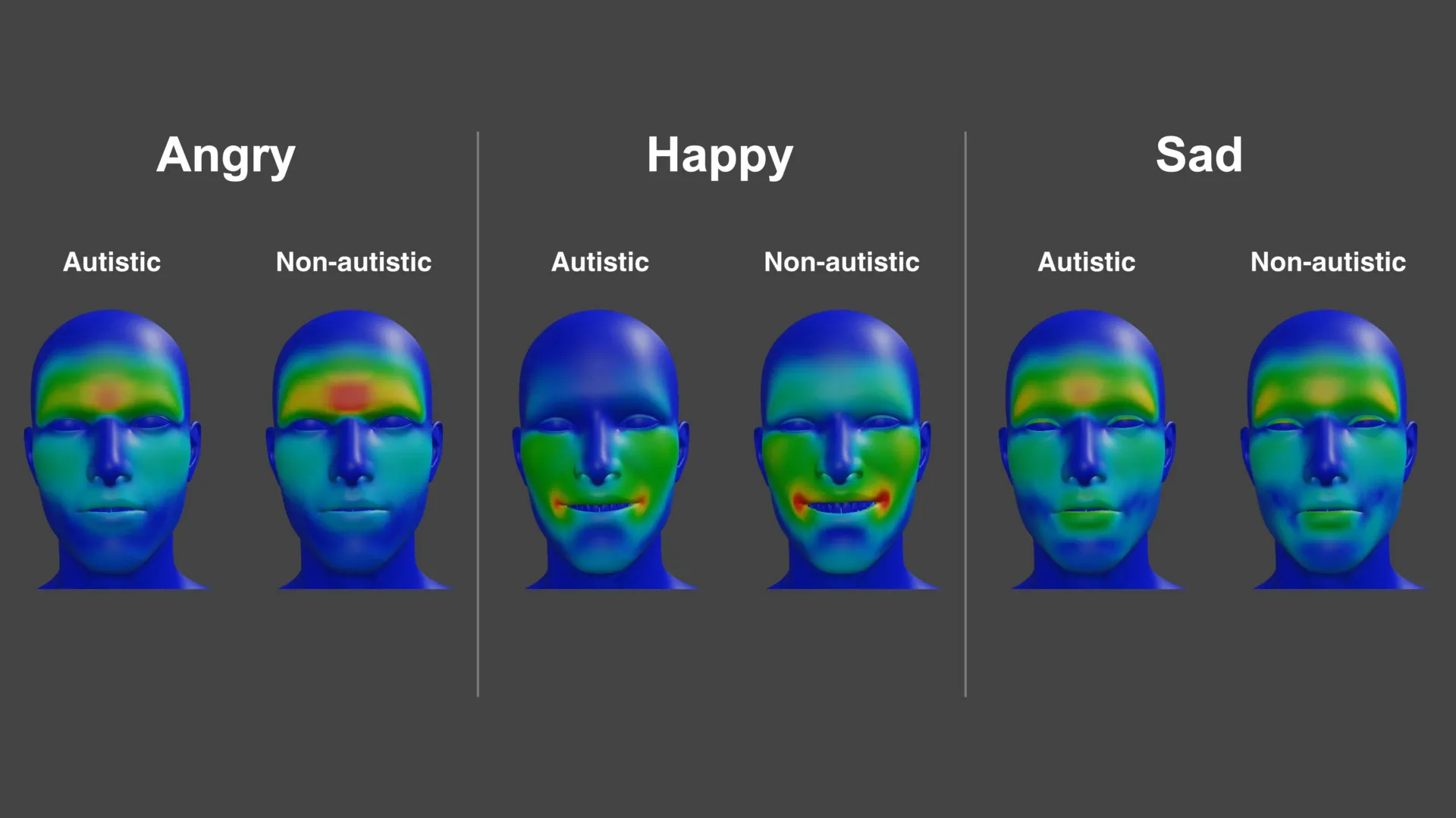
    A new study suggests autistic and non-autistic adults can express the same basic emotions with different facial movement patterns, a gap that may contribute to frequent misunderstandings in everyday interactions. Researchers say the findings support a growing view that communication difficulties can be two-way rather than rooted in a lack of emotion.

    Working with participants in both groups, scientists used detailed facial motion tracking to build a high-resolution map of how expressions for emotions such as anger, happiness and sadness are produced. The project generated more than 265 million data points, creating a large library of facial movements designed to capture subtle differences in how expressions form.

    How the researchers measured expressions

    The study involved 25 autistic adults and 26 non-autistic adults, who together produced close to 5 000 expressions under different conditions. Participants were asked to display emotions while matching facial movements to sounds and while speaking, allowing the team to examine how expressions change across contexts.

    Across tasks, autistic participants showed a wider range of unique expression patterns than their non-autistic peers, the researchers reported. For anger, the autistic group relied more on mouth movement and less on eyebrow movement, while happiness tended to be expressed with a subtler smile and sadness showed a different configuration around the upper lip.

    Alexithymia adds another layer

    The team also examined alexithymia, a trait involving difficulty identifying and describing one’s own emotions that is more common in autism than in the general population. Higher levels of alexithymia were associated with less clearly defined expressions for anger and happiness, which can make the displayed emotion appear more ambiguous.

    Lead researcher Connor Keating said the differences were not only about what expressions look like, but also how smoothly they are formed over time. Senior author Jennifer Cook argued the results fit a communication mismatch model, where both autistic and non-autistic people can misinterpret each other’s emotional signals.

    The research was published in Autism Research and was supported by the UK Medical Research Council and the EU Horizon 2020 programme. The authors said the growing dataset could help improve future studies of emotion recognition, as well as training approaches aimed at reducing everyday misreadings.

  • Depression research takes a new turn: Scientists trace the disorder to specific brain cell types

    Depression research takes a new turn: Scientists trace the disorder to specific brain cell types

    Scientists at McGill University and the Douglas Research Centre have identified specific brain cell types that show altered activity in people with major depression, a finding that could help sharpen the search for more targeted treatments.

    The work, published in Nature Genetics, combines genetic risk signals with cell-level measurements from human brain tissue to pinpoint where depression-related changes appear most strongly.

    Depression is among the world’s leading causes of disability, and many patients do not respond fully to existing therapies. Researchers have long suspected that depression involves measurable biological changes, but mapping them to precise cell types has been difficult.

    Rare brain tissue, sharper tools

    The team relied on post-mortem samples from the Douglas-Bell Canada Brain Bank, one of the specialized collections that includes tissue from people diagnosed with psychiatric conditions.

    Using single-nucleus methods, the researchers profiled gene regulation and gene activity across thousands of individual cells, comparing samples from 59 people with depression and 41 without it.

    Neurons and microglia stand out

    The analysis highlighted two cell populations with notable differences in depression: a group of excitatory neurons involved in mood and stress-related circuits, and a subtype of microglia, immune cells that help regulate inflammation in the brain.

    In both cell types, multiple genes showed altered patterns of activity, suggesting that disruptions in neural signaling and immune-related pathways may converge in the disorder.

    What this could mean for treatment

    By tying depression-associated genetic mechanisms to defined cell types, the study offers a clearer roadmap for experiments that test how these cellular shifts affect brain function over time.

    Researchers caution that the findings do not translate directly into an immediate new therapy, but they may help guide drug development and biomarker research toward more precise biological targets.

    Senior author Gustavo Turecki said the approach provides a clearer picture of where disruptions occur and which cells are involved, reinforcing the view that depression reflects identifiable brain changes rather than a purely psychological experience.