Blog

  • Autism facial expressions mapped in vast new dataset, offering clues to why emotions are often misread

    Autism facial expressions mapped in vast new dataset, offering clues to why emotions are often misread

    A new study suggests autistic and non-autistic adults can express the same basic emotions with different facial movement patterns, a gap that may contribute to frequent misunderstandings in everyday interactions. Researchers say the findings support a growing view that communication difficulties can be two-way rather than rooted in a lack of emotion.

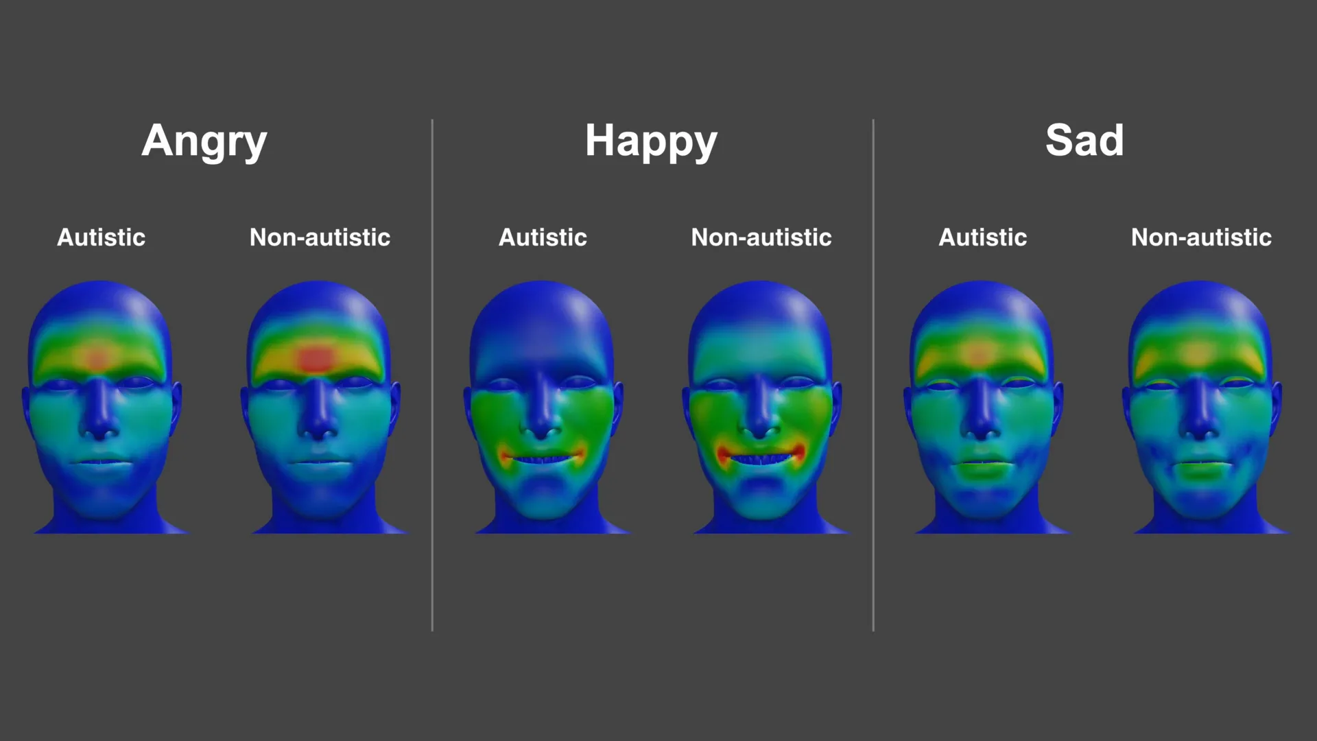
    Working with participants in both groups, scientists used detailed facial motion tracking to build a high-resolution map of how expressions for emotions such as anger, happiness and sadness are produced. The project generated more than 265 million data points, creating a large library of facial movements designed to capture subtle differences in how expressions form.

    How the researchers measured expressions

    The study involved 25 autistic adults and 26 non-autistic adults, who together produced close to 5 000 expressions under different conditions. Participants were asked to display emotions while matching facial movements to sounds and while speaking, allowing the team to examine how expressions change across contexts.

    Across tasks, autistic participants showed a wider range of unique expression patterns than their non-autistic peers, the researchers reported. For anger, the autistic group relied more on mouth movement and less on eyebrow movement, while happiness tended to be expressed with a subtler smile and sadness showed a different configuration around the upper lip.

    Alexithymia adds another layer

    The team also examined alexithymia, a trait involving difficulty identifying and describing one’s own emotions that is more common in autism than in the general population. Higher levels of alexithymia were associated with less clearly defined expressions for anger and happiness, which can make the displayed emotion appear more ambiguous.

    Lead researcher Connor Keating said the differences were not only about what expressions look like, but also how smoothly they are formed over time. Senior author Jennifer Cook argued the results fit a communication mismatch model, where both autistic and non-autistic people can misinterpret each other’s emotional signals.

    The research was published in Autism Research and was supported by the UK Medical Research Council and the EU Horizon 2020 programme. The authors said the growing dataset could help improve future studies of emotion recognition, as well as training approaches aimed at reducing everyday misreadings.

  • Depression research takes a new turn: Scientists trace the disorder to specific brain cell types

    Depression research takes a new turn: Scientists trace the disorder to specific brain cell types

    Scientists at McGill University and the Douglas Research Centre have identified specific brain cell types that show altered activity in people with major depression, a finding that could help sharpen the search for more targeted treatments.

    The work, published in Nature Genetics, combines genetic risk signals with cell-level measurements from human brain tissue to pinpoint where depression-related changes appear most strongly.

    Depression is among the world’s leading causes of disability, and many patients do not respond fully to existing therapies. Researchers have long suspected that depression involves measurable biological changes, but mapping them to precise cell types has been difficult.

    Rare brain tissue, sharper tools

    The team relied on post-mortem samples from the Douglas-Bell Canada Brain Bank, one of the specialized collections that includes tissue from people diagnosed with psychiatric conditions.

    Using single-nucleus methods, the researchers profiled gene regulation and gene activity across thousands of individual cells, comparing samples from 59 people with depression and 41 without it.

    Neurons and microglia stand out

    The analysis highlighted two cell populations with notable differences in depression: a group of excitatory neurons involved in mood and stress-related circuits, and a subtype of microglia, immune cells that help regulate inflammation in the brain.

    In both cell types, multiple genes showed altered patterns of activity, suggesting that disruptions in neural signaling and immune-related pathways may converge in the disorder.

    What this could mean for treatment

    By tying depression-associated genetic mechanisms to defined cell types, the study offers a clearer roadmap for experiments that test how these cellular shifts affect brain function over time.

    Researchers caution that the findings do not translate directly into an immediate new therapy, but they may help guide drug development and biomarker research toward more precise biological targets.

    Senior author Gustavo Turecki said the approach provides a clearer picture of where disruptions occur and which cells are involved, reinforcing the view that depression reflects identifiable brain changes rather than a purely psychological experience.

  • Study finds the brain processes speech in AI-like layers, offering new clues to how meaning is built

    Study finds the brain processes speech in AI-like layers, offering new clues to how meaning is built

    New research suggests the human brain may understand spoken language through a layered, step-by-step process that closely parallels how large language models handle text. By tracking neural activity as people listened to a continuous story, scientists found patterns that align with the progression from simpler to more complex representations seen in modern AI.

    The work, published in Nature Communications, analyzed high-temporal-resolution recordings from electrodes placed on the brain surface in clinical settings. Researchers compared the timing of neural responses with internal representations from well-known language models, including GPT-2 and Meta’s Llama 2.

    How meaning appears to unfold

    The team reports that early brain signals corresponded more closely to the earlier computational stages of AI systems that focus on basic word-level features. Later neural responses matched deeper model layers that integrate broader context, linking words into higher-level meaning.

    This alignment was especially pronounced in established language regions, including areas often associated with speech production and comprehension such as Broca’s area. In these regions, the strongest match tended to appear later in time, consistent with a gradual buildup of meaning.

    Rethinking classic language theories

    The findings add weight to the idea that comprehension is not driven primarily by rigid, rule-based structures applied instantly to each sentence. Instead, the results support a view in which the brain continuously updates interpretations as more context arrives, resembling statistical inference more than fixed symbolic parsing.

    Researchers also evaluated traditional linguistic descriptors, such as phoneme- and morpheme-level features, and found they explained real-time neural activity less effectively than the contextual features derived from AI models. That gap, the authors argue, suggests that context-rich representations may better capture how the brain tracks meaning in natural speech.

    A dataset meant to accelerate research

    Alongside the paper, the team released a public dataset designed to help other labs test competing theories of language processing against neural measurements. By pairing brain recordings with model-derived language features, the resource is intended to make comparisons across studies more consistent and reproducible.

    Experts caution that similarities do not mean the brain works the same way as today’s AI, which is trained on vast text corpora and built from artificial neural networks. Still, the results strengthen the case that AI language models can serve as useful scientific tools for probing how the brain constructs meaning over time.

  • Harvard study traces a gut bacteria link to depression, pointing to an environmental chemical that may fuel inflammation

    Harvard study traces a gut bacteria link to depression, pointing to an environmental chemical that may fuel inflammation

    Harvard Medical School researchers have outlined a new molecular pathway that may help explain why certain gut bacteria are repeatedly associated with major depressive disorder. The work centers on Morganella morganii and an immune reaction that could connect gut chemistry to brain health.

    In the study, scientists found that an environmental contaminant called diethanolamine, or DEA, can be incorporated into a lipid-like molecule produced by M. morganii in the gut. That altered molecule appears to behave differently than the standard version, shifting from relatively inert to immune-activating.

    A chemical swap with immune effects

    Laboratory tests showed the modified bacterial molecule can stimulate inflammatory signaling, including elevated release of cytokines such as interleukin-6, or IL-6. IL-6 has been widely studied as part of the broader link between inflammation and depressive symptoms in some patients.

    The authors argue this provides a clearer mechanism than earlier correlation-based microbiome findings, though it does not prove the pathway causes depression in humans. They say additional clinical work is needed to determine how often this chemistry occurs in real-world conditions and who may be most affected.

    Why inflammation matters in depression

    Inflammation has long been investigated as one contributor to depression, with evidence suggesting a subset of cases may involve immune dysregulation. The new results fit into that view by describing how a bacterial product, altered by exposure to a common industrial chemical, might intensify inflammatory signaling.

    The team also notes that M. morganii has been associated in prior research with inflammatory conditions beyond depression, including metabolic and gastrointestinal diseases. That pattern, they say, supports the idea that the bacterium’s metabolites can interact with the immune system in clinically meaningful ways.

    Potential paths for diagnosis and treatment

    The researchers suggest DEA-related chemistry could eventually help identify biological subtypes of depression, potentially using exposure markers or microbial metabolites as part of a diagnostic approach. Any such use would require validation in human cohorts and careful separation of correlation from causation.

    More broadly, the findings add momentum to efforts exploring treatments that target inflammation for selected patients, alongside established psychological and pharmacological therapies. The authors describe their work as a framework for scanning other gut microbes for similar pollutant-driven metabolic changes.

    The study was published in the Journal of the American Chemical Society and combined expertise in bacterial small-molecule chemistry and microbiome-immunity interactions. Researchers involved also highlighted the role of cross-disciplinary microbiome research in moving from broad associations toward specific, testable mechanisms.

  • Brain stimulation linked to small boost in generosity: What a new PLOS Biology study found

    Brain stimulation linked to small boost in generosity: What a new PLOS Biology study found

    Non-invasive brain stimulation that nudges two brain regions to operate in sync may slightly increase generous choices, according to a study published in PLOS Biology on February 10. Researchers say the results add evidence that specific brain-network communication can shape social decision-making.

    The international team, led by Jie Hu of East China Normal University with collaborators at the University of Zurich, tested whether coordinating activity between frontal and parietal areas affects altruism. These regions are often associated with goal-directed behavior and higher-level reasoning during complex choices.

    Testing generosity in a lab game

    The experiment included 44 participants who completed 540 rounds of a standard behavioral task known as the Dictator Game. In each round, a participant decided how to split money with another person, with the amounts varying across decisions.

    During the task, the researchers applied transcranial alternating current stimulation, a technique designed to influence brain rhythms through weak electrical currents delivered via the scalp. The goal was to encourage synchrony between the targeted frontal and parietal regions at specific oscillation frequencies.

    Gamma synchrony showed the clearest shift

    When stimulation was set to strengthen gamma-band synchrony between the two regions, participants became modestly more likely to choose larger splits for the other person. The effect was most apparent even in situations where giving more meant the participant would take less than their counterpart.

    Using computational modeling, the team reported that the stimulation appeared to change how people weighed outcomes, increasing the importance placed on the other person’s payoff. The authors described this as a measurable shift in value computations rather than a simple preference for equal splits.

    Limits and what comes next

    The researchers noted they did not directly record neural activity during stimulation, meaning the intended brain synchrony was inferred rather than confirmed in real time. They suggested future studies combining stimulation with EEG could verify how brain signals change and how long any behavioral effects last.

    Coauthor Christian Ruff said the work helps link a specific communication pattern between brain regions to altruistic choices, while Hu emphasized the study’s attempt to demonstrate cause and effect. The team cautioned that the increase in generosity was small and the findings do not imply a tool for controlling behavior outside controlled research settings.

  • Fish oil and brain injury recovery: Study flags EPA as a potential risk in repeated concussions

    Fish oil and brain injury recovery: Study flags EPA as a potential risk in repeated concussions

    A new study from the Medical University of South Carolina is raising fresh concerns about fish oil supplements, especially for people who experience repeated mild traumatic brain injuries. Writing in the journal Cell Reports, researchers report that these widely used supplements, often promoted as protective for the brain, could actually interfere with healing after injury.

    The research was led by neuroscientist Onder Albayram, Ph.D., an associate professor at MUSC and a member of the National Trauma Society Committee. His team focused on the biological processes involved in repairing blood vessels in the brain after injury.

    Rising Popularity of Omega-3 Supplements

    Interest in omega-3 fatty acids, the key components of fish oil, has been growing rapidly. According to Fortune Business Insights, these supplements are now appearing not only in capsules but also in drinks, dairy alternatives, and snack products.

    That surge in popularity does not surprise Albayram. “Fish oil supplements are everywhere, and people take them for a range of reasons, often without a clear understanding of their long-term effects,” he said.

    “But in terms of neuroscience, we still don’t know whether the brain has resilience or resistance to this supplement. That’s why ours is the first such study in the field.”

    Albayram collaborated with Eda Karakaya, Ph.D., Adviye Ergul, M.D., Ph.D., and several other researchers at MUSC and partner institutions. Among them was Semir Beyaz, Ph.D., at the Cold Spring Harbor Laboratory Cancer Center in New York.

    EPA Identified as a Potential Weak Point in Brain Recovery

    The team discovered what they describe as a context-dependent metabolic vulnerability. In simple terms, this means that changes in how cells use energy may reduce the brain’s ability to recover under certain conditions. This vulnerability appears to be linked to the buildup of eicosapentaenoic acid, or EPA, one of the main omega-3 fatty acids found in fish oil.

    In their experimental models, higher levels of EPA in the brain were associated with weaker repair after injury.

    Albayram noted that not all omega-3s behave the same way. Docosahexaenoic acid, or DHA, is well known for its beneficial role in the brain and is a major part of neuronal membranes. EPA, however, follows a different pathway. It is less incorporated into brain structures, and its effects can vary depending on how long it is present and the surrounding biological conditions. Because of this, the long-term impact of omega-3 intake on brain recovery and blood vessel adaptation has remained unclear.

    Experiments Link Diet, Brain Biology, and Recovery

    To better understand these effects, the researchers used a series of models to connect diet, brain function, and healing. In mice, they examined how long-term fish oil use influenced the brain’s response to repeated mild head impacts. Their focus was on signals related to blood vessel stability and repair.

    They also studied human brain microvascular endothelial cells, which form part of the barrier between the brain and the bloodstream. In these cells, EPA, but not DHA, was linked to reduced repair capacity, aligning with the findings from the animal models.

    To extend the findings to real-world disease, the team analyzed postmortem brain tissue from individuals diagnosed with chronic traumatic encephalopathy (CTE) who had a history of repeated brain injury.

    The researchers described the results as having “implications for precision nutrition, therapeutic strategies and the design of dietary interventions targeting brain injury and neurodegeneration.”

    Key Findings From the Study

    The study identified several major patterns, which are summarized below along with simplified explanations.

    1. EPA-driven neurovascular instability triggers perivascular tauopathy and cognitive decline following TBI.

    “In a sensitive brain state modeled in mice, long-term fish oil supplementation revealed a delayed vulnerability. The animals showed poorer neurological and spatial learning performance over time, together with clear evidence of vascular-associated tau accumulation in the cortex, linking impaired recovery to neurovascular dysfunction and perivascular tau pathology,” Albayram said.

    1. EPA reprograms cortical transcriptional responses and suppresses angiogenic signaling following traumatic brain injury.

    “In the injured cortex, the team observed a coordinated shift in gene programs that normally support vascular stability and repair,” Albayram said. “The pattern included reduced expression of genes tied to extracellular matrix organization and endothelial integrity, alongside broader changes consistent with altered lipid handling after injury.”

    1. EPA utilization under permissive metabolic conditions impairs angiogenesis and endothelial integrity, recapitulating post-traumatic brain injury cerebrovascular dysfunction.

    Albayram said that in human brain microvascular endothelial cells, EPA did not act as a universal toxin. “Instead, when cells were placed in conditions that encouraged fatty acid engagement, EPA was associated with weaker angiogenic network formation and reduced endothelial barrier integrity, matching key features of the neurovascular repair deficit seen in vivo.”

    1. CTE brain reveals neurovascular and fatty acid metabolic reprogramming consistent with EPA-linked vulnerability.

    “In postmortem cortex from neuropathologically confirmed CTE cases with a history of repetitive brain injury, the researchers found evidence of disrupted fatty acid balance and broad transcriptional changes affecting vascular and metabolic pathways,” Albayram said. “This human arm was used to provide translational context, asking whether chronic disease tissue shows convergent signatures of altered lipid handling and reduced vascular stability.”

    What the Findings Mean for Fish Oil Use

    Albayram stressed that the study should not be interpreted as a blanket warning against fish oil. “I am not saying fish oil is good or bad in some universal way,” he said. “What our data highlight is that biology is context-dependent. We need to understand how these supplements behave in the body over time, rather than assuming the same effect applies to everyone.”

    The researchers hope their work encourages a more careful look at omega-3 supplementation, both in clinical settings and among the general public. Their experiments focused on a specific scenario, repeated mild brain injury, and used CTE tissue to provide supporting observations rather than direct proof of cause and effect.

    “As with any study, there are important boundaries,” Albayram said. “In the human CTE tissue, we can observe patterns, but we cannot prove what drove them. We also cannot capture every variable that shapes omega-3 handling in real life, including overall diet, health status and lifestyle.”

    Next Steps in Understanding Omega-3 Effects

    The team plans to continue investigating how EPA moves through the body, including how it is absorbed, transported, and distributed. They are especially interested in the mechanisms that control fatty acid movement.

    “This paper is a starting point,” Albayram said, “but it is an important one. It opens a new conversation about precision nutrition in neuroscience, and it gives the field a framework to ask better, more testable questions.”
  • University of Illinois study suggests joint savoring can strengthen relationships, especially under stress

    University of Illinois study suggests joint savoring can strengthen relationships, especially under stress

    Couples who deliberately pause to appreciate enjoyable moments together tend to report stronger, more stable relationships, according to new research from the University of Illinois Urbana-Champaign. The findings suggest that this shared habit, known as joint savoring, is linked to higher satisfaction and fewer conflicts.

    Savoring is commonly described by psychologists as slowing down to notice positive experiences and letting them register, whether in the present or through remembering the past and anticipating the future. While earlier work has tied savoring to individual well-being, the Illinois team examined what changes when partners do it together.

    How joint savoring was measured

    The study analyzed survey responses from 589 adults across the United States who were in committed relationships, most of them married. Participants were asked how often they and their partners intentionally focused on positive shared experiences, using a relationship-focused measure adapted from established savoring research.

    Respondents also reported on relationship quality, including satisfaction, communication conflict, and confidence that the relationship would last. They answered separate questions about stress and well-being, capturing whether they felt on top of responsibilities or overwhelmed in the prior month.

    A buffer when stress increases

    Researchers found that people who reported more joint savoring also reported less conflict, greater relationship satisfaction, and stronger belief in their shared future. The same group also showed signs of better personal well-being, indicating potential spillover benefits beyond the relationship itself.

    The pattern was most pronounced among people experiencing higher stress, where joint savoring appeared to function as a protective factor. In other words, when pressures rose, couples who routinely focused on positive shared moments were more likely to maintain confidence in their relationship and protect mental health.

    Why small rituals may matter

    The authors argue that joint savoring is practical because it does not require major life changes, only intentional attention. They suggest couples can build it into normal routines, such as talking through a good memory, lingering over a meal, or planning something enjoyable together.

    The research also underscores a limitation common to survey-based studies: the data reflects self-reports rather than observations of both partners in real time. Still, the findings add to a growing body of evidence that simple, repeatable relationship habits can help couples stay resilient, particularly during stressful periods.

  • Rutgers study suggests internalized stress could speed memory decline in older Chinese Americans

    Rutgers study suggests internalized stress could speed memory decline in older Chinese Americans

    Stress that people keep to themselves may be quietly linked to faster memory decline in older Chinese Americans, according to new research from Rutgers Health. The findings add detail to how emotional strain can shape brain aging in a population often underrepresented in dementia studies.

    The study, published in The Journal of Prevention of Alzheimer’s Disease, analyzed how social and behavioral factors relate to changes in memory over time. Researchers focused on Chinese adults aged 60 and older, noting that more tailored evidence is needed as the older Asian American population grows.

    What internalized stress looks like

    In the research, internalized stress referred to turning distress inward, including feelings such as hopelessness and absorbing stressful experiences rather than expressing or resolving them. The team reported that this pattern showed the clearest association with worsening memory across multiple assessments.

    The authors also pointed to cultural and life circumstances that can intensify hidden strain, including pressure to appear resilient and the practical stressors many older immigrants face. Language barriers, social isolation and adapting to different norms can compound stress over time.

    Evidence drawn from a major cohort

    To examine long-term patterns, the researchers used data from the Population Study of ChINese Elderly, a large community-based cohort in the Chicago area. The dataset followed more than 1 500 participants through interviews conducted from 2011 to 2017.

    Alongside internalized stress, the team evaluated neighborhood cohesion and external stress relief factors. Those measures did not show the same clear relationship with memory changes as internalized stress did, underscoring the potential importance of how stress is processed emotionally.

    Why the findings matter now

    The researchers emphasized that internalized stress is potentially modifiable, which may open doors for prevention-focused support. They argued that culturally sensitive approaches could help identify distress that is easy to miss and may influence cognitive health.

    While the study does not prove that stress causes dementia, it strengthens the case that emotional well-being and brain health are closely connected. The authors said the results could inform community programs and clinical screening that better fit the experiences of aging Chinese American adults.

  • New research explains why human language resists code-like efficiency and what it means for AI

    New research explains why human language resists code-like efficiency and what it means for AI

    Human language can seem inefficient compared with computer code, but researchers argue that its structure is optimized for the brain rather than for maximum compression. A new modeling study suggests people rely on familiar patterns to reduce mental effort during real-time conversation.

    The work, by linguist Michael Hahn and cognitive scientist Richard Futrell, was published in Nature Human Behaviour. Using information-theory-based modeling, they examined why languages worldwide tend to favor predictable word patterns instead of highly compact encodings.

    Efficiency for brains, not bits

    In principle, the same message could be transmitted with fewer symbols, similar to how computers use binary strings. The researchers contend that such a system would be harder for humans to learn and process because it would not align with how people store knowledge and anticipate meaning.

    Natural language, they argue, is tightly linked to shared experience, letting listeners map words onto familiar concepts quickly. That connection helps speakers avoid creating arbitrary, maximally compressed labels that would be information-dense but difficult to interpret.

    Predictability lowers cognitive load

    The model emphasizes that comprehension is incremental: listeners use each word to narrow down likely meanings before a sentence ends. This predictive processing makes everyday communication feel almost automatic, even if it is not mathematically optimal in terms of compression.

    As an illustration, the authors point to how grammatical order guides expectations in languages such as German. When familiar cues arrive in the expected sequence, the brain can prune unlikely interpretations early, whereas scrambled word orders force more effortful processing.

    What the findings suggest for AI

    The researchers say the results help explain why languages converge on structures that are learnable and robust under noisy, fast conditions like speech. Rather than chasing minimal code length, languages appear to balance expressiveness with the constraints of memory, attention, and prediction.

    The same logic could inform how developers evaluate and design large language models, which already rely heavily on predicting likely next words. The study suggests that systems built to communicate smoothly with people may benefit from prioritizing human-friendly predictability over pure information compression.

  • New brain imaging study suggests intelligence hinges on whole-brain network efficiency, not a single region

    New brain imaging study suggests intelligence hinges on whole-brain network efficiency, not a single region

    Modern neuroscience often describes the brain as a collection of specialized systems. Functions such as attention, perception, memory, language, and reasoning have each been linked to specific brain networks, and scientists have typically studied these systems separately.

    This approach has produced major breakthroughs. However, it has not fully explained a central feature of human thinking: how all these separate systems come together to form a single, unified mind.

    Researchers at the University of Notre Dame set out to address that question. Using advanced neuroimaging, they examined how the brain is organized overall and how that organization gives rise to intelligence.

    “Neuroscience has been very successful at explaining what particular networks do, but much less successful at explaining how a single, coherent mind emerges from their interaction,” said Aron Barbey, the Andrew J. McKenna Family Professor of Psychology in Notre Dame’s Department of Psychology.

    General Intelligence and Connected Cognitive Abilities

    Psychologists have long observed that skills like attention, memory, perception, and language tend to be linked. People who perform well in one area often perform well in others. This pattern is known as “general intelligence.” It influences how effectively individuals learn, solve problems, and adapt across academic, professional, social, and health settings.

    For more than a century, this pattern has suggested that human cognition is unified at a deep level. What scientists have lacked is a clear explanation for why that unity exists.

    “The problem of intelligence is not one of functional localization,” said Barbey, who also directs the Notre Dame Human Neuroimaging Center and the Decision Neuroscience Laboratory. “Contemporary research often asks where general intelligence originates in the brain — focusing primarily on a specific network of regions within the frontal and parietal cortex. But the more fundamental question is how intelligence emerges from the principles that govern global brain function — how distributed networks communicate and collectively process information.”

    To explore this broader perspective, Barbey and his team, including lead author and Notre Dame graduate student Ramsey Wilcox, tested a framework known as the Network Neuroscience Theory. Their findings were published in Nature Communications.

    The Network Neuroscience Theory Explained

    According to the researchers, general intelligence is not a specific ability or mental strategy. Instead, it reflects a pattern in which many cognitive skills are positively related. They propose that this pattern stems from how efficiently the brain’s networks are structured and how well they work together.

    To evaluate this idea, the team analyzed brain imaging and cognitive performance data from 831 adults in the Human Connectome Project. They also examined an independent group of 145 adults in the INSIGHT Study, funded by the Intelligence Advanced Research Projects Activity’s SHARP program. By combining measures of brain structure and brain function, the researchers created a detailed picture of large-scale brain organization.

    Rather than tying intelligence to a single brain region or function, the Network Neuroscience Theory views it as a property of the brain as a whole. Intelligence, in this framework, depends on how effectively networks coordinate and reorganize themselves to handle different challenges.

    Barbey and Wilcox describe this as a major shift in perspective.

    “We found evidence for system-wide coordination in the brain that is both robust and adaptable,” Wilcox said. “This coordination does not carry out cognition itself, but determines the range of cognitive operations the system can support.”

    “Within this framework, the brain is modeled as a network whose behavior is constrained by global properties such as efficiency, flexibility and integration,” Wilcox said. “These properties are not tied to individual tasks or brain networks, but are characteristics of the system as a whole, shaping every cognitive operation without being reducible to any one of them.”

    “Once the question shifts from where intelligence is to how the system is organized,” Wilcox noted, “the empirical targets change.”

    Intelligence as Whole Brain Coordination

    The findings supported four main predictions of the Network Neuroscience Theory.

    First, intelligence does not reside in a single network. It arises from processing distributed across many networks. The brain must divide tasks among specialized systems and combine their outputs when necessary.

    Second, successful coordination requires strong integration and long-distance communication. Barbey described “a large and complex system of connections that serve as ‘shortcuts’ linking distant brain regions and integrating information across the networks.” These connections allow far apart areas of the brain to exchange information efficiently, supporting unified processing.

    Third, integration depends on regulatory regions that guide how information flows. These hubs help orchestrate activity across networks, selecting the right systems for the job. Whether someone is interpreting subtle clues, learning a new skill, or deciding between careful analysis and quick intuition, these regulatory areas help manage the process.

    Finally, general intelligence depends on balancing local specialization with global integration. The brain performs best when tightly connected local clusters operate efficiently while still maintaining short communication paths to distant regions. This balance supports flexible and effective problem solving.

    Across both groups studied, differences in general intelligence consistently matched these large-scale organizational features. No single brain area or traditional “intelligence network” explained the results.

    “General intelligence becomes visible when cognition is coordinated,” Barbey noted, “when many processes must work together under system-level constraints.”

    Implications for Artificial Intelligence and Brain Development

    The implications extend beyond understanding human intelligence. By focusing on large-scale brain organization, the findings offer insight into why the mind functions as a unified system in the first place.

    This perspective may also explain why intelligence tends to increase during childhood, decline with aging, and be especially vulnerable to widespread brain injury. In each situation, what changes most is large-scale coordination rather than isolated functions.

    The results also contribute to debates about artificial intelligence. If human intelligence depends on system-level organization rather than a single general-purpose mechanism, then building artificial general intelligence may require more than simply scaling up specialized tools.

    “This research can push us into thinking about how to use design characteristics of the human brain to motivate advances in human-centered, biologically inspired artificial intelligence,” Barbey said.

    “Many AI systems can perform specific tasks very well, but they still struggle to apply what they know across different situations.” Barbey said. “Human intelligence is defined by this flexibility — and it reflects the unique organization of the human brain.”

    The research was conducted with co-authors Babak Hemmatian and Lav Varshney of Stony Brook University.