Blog

  • Study suggests a part of the human auditory cortex is uniquely tuned to chimpanzee calls

    Study suggests a part of the human auditory cortex is uniquely tuned to chimpanzee calls

    The human brain is not limited to recognizing our own voices. Research from the University of Geneva (UNIGE) has revealed that specific parts of the auditory cortex react strongly to chimpanzee vocalizations. Chimpanzees are our closest relatives both genetically and in the types of sounds they produce. The study, which appears in the journal eLife, indicates that certain subregions of the brain may be especially tuned to the calls of particular primate species. This insight offers a new way to explore how voice recognition emerged and how it may relate to the development of language.

    Human voices play a central role in social communication, and a significant portion of the auditory cortex is devoted to interpreting them. Researchers wanted to know whether these abilities have deeper evolutionary origins. To investigate this question, scientists from UNIGE’s Faculty of Psychology and Educational Sciences used a comparative approach grounded in species evolution. By examining how the human brain processes the vocalizations of closely related species, such as chimpanzees, bonobos and macaques, they aimed to identify traits shared with other primates. This approach helps shed light on how the neural foundations of vocal communication began to emerge long before language existed.

    Studying How the Brain Responds to Primate Calls

    In the experiment, 23 human volunteers listened to vocal sounds from four species. Human voices served as the control group. Chimpanzee calls were included because these primates are closely related to us both genetically and acoustically. Bonobo vocalizations were also tested, even though they often sound more like birdsong. Macaque calls were added because these primates are more distantly related to humans both evolutionarily and acoustically. Researchers used functional magnetic resonance imaging (fMRI) to examine activity across the auditory cortex. “Our intention was to verify whether a subregion sensitive specifically to primate vocalizations existed,” explains Leonardo Ceravolo, research associate at UNIGE’s Faculty of Psychology and Educational Sciences and first author of the study.

    A Distinct Neural Response to Chimpanzee Vocalizations

    The results confirmed their expectations. A part of the auditory cortex known as the superior temporal gyrus, which plays a key role in processing sounds related to language, music and emotional cues, showed increased activation when participants heard certain primate calls. “When participants heard chimpanzee vocalizations, this response was clearly distinct from that triggered by bonobos or macaques.”

    This pattern is particularly striking because bonobos are just as genetically close to humans as chimpanzees, yet their vocalizations differ greatly in acoustic structure. The findings suggest that both evolutionary closeness and similarity in sound features influence how the human brain reacts.

    What the Findings Suggest About Language Evolution

    This discovery offers new directions for understanding how the neural basis of communication evolved. It indicates that some parts of the human brain may have preserved a sensitivity to the calls of our closest primate relatives. “We already knew that certain areas of the animal brain reacted specifically to the voices of their fellow creatures. But here, we show that a region of the adult human brain, the anterior superior temporal gyrus, is also sensitive to non-human vocalizations,” notes Leonardo Ceravolo.

    These results support the idea that humans and great apes share vocal processing abilities that existed before spoken language emerged. They may also help explain how voice recognition develops in early life. For instance, this line of research could clarify how babies begin recognizing familiar voices while still in utero.

  • MIT study links GRIN2A mutation to slower reality updating in schizophrenia, pointing to a treatable brain circuit

    MIT study links GRIN2A mutation to slower reality updating in schizophrenia, pointing to a treatable brain circuit

    Researchers at MIT report that a mutation in the gene GRIN2A may interfere with how the brain updates beliefs when new information arrives, a cognitive difficulty often seen in schizophrenia. In mouse experiments, the change was tied to slower, less adaptive decision-making in a shifting environment.

    Schizophrenia affects about 1% of people and has a strong genetic component, though the biology connecting risk genes to symptoms has been hard to pin down. Large genomic studies have identified many associated variants, but many sit in non-coding DNA, making their functional impact difficult to interpret.

    From genetic signal to mechanism

    To narrow in on mutations that directly alter proteins, the team drew on large-scale exome sequencing that compares protein-coding regions across people with schizophrenia and unaffected controls. That work has helped highlight a smaller set of genes where rare disruptive mutations can substantially increase risk.

    GRIN2A stands out because it encodes a subunit of the NMDA receptor, a key component of glutamatergic signaling involved in learning, plasticity and cognitive control. NMDA receptor dysfunction has long been considered relevant to schizophrenia, but linking specific mutations to circuit-level effects has remained challenging.

    Decision task reveals slower adaptation

    In the study’s behavioral task, mice chose between two levers with different reward sizes and different effort costs that changed over time. Typical mice shifted to the more efficient option once the higher-reward choice became too costly, reflecting flexible updating as conditions evolved.

    Mice carrying the GRIN2A-related mutation took longer to commit, continuing to alternate between choices after the balance of effort and reward had effectively changed. The researchers interpret the pattern as reduced ability to incorporate new evidence quickly, leaving prior expectations to dominate behavior for longer.

    A circuit that can be nudged

    Brain measurements pointed to altered activity in the mediodorsal thalamus and its connections with the prefrontal cortex, a pathway central to executive function and decision-making. The team reports that this thalamocortical circuit appeared to represent changing option values differently in the mutated mice.

    Using optogenetics to activate neurons in the mediodorsal thalamus, the researchers were able to push behavior toward the more adaptive pattern seen in control animals. While only a subset of patients would be expected to carry GRIN2A mutations, the results suggest the same circuit could contribute to cognitive symptoms across broader groups.

    The authors frame the work as a step toward treatments that target cognition, an area where many patients continue to experience impairment even when hallucinations or delusions are reduced. Next efforts focus on identifying druggable nodes in the thalamus–prefrontal pathway that might restore more flexible updating without invasive methods.

  • UC Berkeley study points to oxytocin as the fast track to friendship, and why some bonds take longer to form

    UC Berkeley study points to oxytocin as the fast track to friendship, and why some bonds take longer to form

    New research from the University of California, Berkeley suggests the hormone oxytocin helps speed up the early stages of friendship formation, sharpening the sense of preferring a familiar peer over a stranger. The work, published in Current Biology, adds nuance to oxytocin’s popular image by focusing on how quickly and selectively social bonds take shape.

    Oxytocin is released during a range of social and bodily experiences, including touch, sex, childbirth and breastfeeding, and it acts in the brain as a neuromodulator. While it is often linked with closeness and trust, scientists have also associated oxytocin signaling with social defensiveness, including stronger in-group and out-group behavior.

    The team studied prairie voles, a species widely used to examine social bonding because individuals form stable, selective relationships. Instead of focusing only on mating pairs, the researchers emphasized peer bonds that resemble human friendships, such as choosing to huddle and groom with one familiar partner rather than spending time with strangers.

    What changed without oxytocin receptors

    Using prairie voles engineered to lack oxytocin receptors, the researchers found the animals were slower to form a peer preference. In tests where typical voles show a strong preference after about 24 hours, the receptor-deficient animals often needed up to a week to reliably choose a familiar partner.

    The difference was not simply that the animals became less social overall. The findings point to reduced selectivity, meaning the altered animals were less consistent about who they sought out and were quicker to lose track of established partners when placed into new group settings.

    Friendship selectivity, not just sociability

    In a mixed-group, multi-room setup designed to mimic a party-like environment, typical voles spent early time near known companions before gradually mingling. Voles without oxytocin receptors mixed more freely from the start, behaving as if prior peer connections carried less weight.

    In another test measuring social motivation, female voles usually worked harder to access a familiar peer than a stranger. The receptor-deficient animals still showed motivation for a mate, but not for a friend, indicating that oxytocin signaling may matter more for the reward value of peer bonds than for mating bonds.

    A new look with oxytocin nanosensors

    To examine whether the brain compensated for missing receptors by releasing more oxytocin, researchers used an oxytocin nanosensor that fluoresces when it detects the molecule. Measurements indicated no excess oxytocin release and, instead, lower release from fewer sites in the nucleus accumbens, a region central to social reward.

    The results help explain why friendships formed more slowly and were less stable in challenging social conditions. Researchers say the work could inform future studies of psychiatric conditions where social bonding is disrupted, while underscoring that oxytocin’s role is complex and context-dependent.

    The study also fits into a growing body of vole research suggesting oxytocin is not strictly required for bonds to exist, but can strongly affect how efficiently they form. By separating friendship-like bonds from mating behavior, the authors argue that the biology of peer relationships deserves attention in its own right.

  • Gut microbiome study points to inflammatory sugars as a potential trigger for ALS and frontotemporal dementia

    Gut microbiome study points to inflammatory sugars as a potential trigger for ALS and frontotemporal dementia

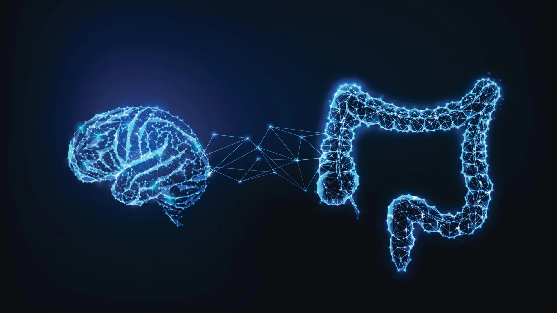
    Researchers at Case Western Reserve University say they have identified a gut-driven immune mechanism that may help explain why brain damage progresses in amyotrophic lateral sclerosis and frontotemporal dementia. The work focuses on specific sugars produced by certain gut bacteria and how they may spark harmful inflammation.

    The study, published in Cell Reports, links bacterial glycogen molecules to immune activation that can injure neurons in models of ALS and FTD. The authors suggest the pathway could become both a diagnostic clue and a target for treatments aimed at the gut rather than the brain alone.

    How the proposed gut pathway works

    According to the researchers, some gut microbes can generate an inflammatory form of glycogen, a complex sugar, that appears to provoke immune responses tied to neurodegeneration. In the study’s patient samples, 70% of 23 people with ALS or FTD showed elevated levels of the implicated bacterial glycogen, compared with about one-third of people without the conditions.

    Aaron Burberry, an assistant professor at Case Western Reserve School of Medicine, said the team found that these microbial sugars can trigger immune reactions that ultimately damage brain cells. The results point to a potential way to interrupt the process by reducing or degrading the inflammatory glycogen.

    Why genes may not be the whole story

    The findings could be especially relevant for carriers of the C9orf72 mutation, the most common known genetic cause of ALS and FTD. Many carriers never develop disease, and the study adds evidence that environmental factors such as the gut microbiome may influence whether symptoms emerge.

    The researchers propose that certain bacterial communities may act as a switch that worsens inflammation in genetically susceptible people. That idea aligns with a growing body of research connecting immune activity, the microbiome, and neurodegenerative disease risk.

    What comes next for treatment research

    In experiments described by the team, lowering levels of the harmful glycogen improved brain health measures and extended lifespan in model systems. Alex Rodriguez-Palacios, also at Case Western, said reducing these sugars was associated with better outcomes, supporting efforts to design therapies that target the compounds in the gut.

    The group plans larger studies to track microbiome changes in ALS and FTD patients, including before and after disease onset where possible. Burberry said the results support clinical trials testing whether glycogen-degrading approaches could slow progression, though timelines and trial designs will depend on additional validation.

  • Study suggests strong social ties may slow biological aging, with epigenetic clocks offering new clues

    Study suggests strong social ties may slow biological aging, with epigenetic clocks offering new clues

    Building strong relationships throughout life — from loving parents in childhood to close friends, active communities, and faith involvement in adulthood — may actually slow how the body ages. Researchers suggest that these “social advantages” can influence biological aging markers known as epigenetic clocks, which track changes in DNA methylation. People who enjoy more supportive and connected lives often appear biologically younger than their chronological age.

    Long-Term Study Links Social Advantage to Youthful Biology

    The findings were published in the October issue of Brain, Behavior and Immunity — Health and draw on data from over 2,100 adults who participated in the long-running Midlife in the United States (MIDUS) study.

    Anthony Ong, a psychology professor at Cornell University, and his colleagues discovered that people with greater “cumulative social advantage” — a measure of lifelong social and emotional support — tended to show slower biological aging and reduced chronic inflammation.

    Measuring the Pace of Aging

    The study examined two leading measures of biological age, called GrimAge and DunedinPACE. Both are epigenetic clocks that scientists use to predict health risks and life expectancy. Participants with richer and more consistent social relationships displayed younger biological profiles on both measures.

    “Cumulative social advantage is really about the depth and breadth of your social connections over a lifetime,” Ong said. “We looked at four key areas: the warmth and support you received from your parents growing up, how connected you feel to your community and neighborhood, your involvement in religious or faith-based communities, and the ongoing emotional support from friends and family.”

    The Biology of Connection

    The researchers hypothesized that sustained social advantage becomes reflected in core regulatory systems linked to aging, including epigenetic, inflammatory and neuroendocrine pathways. Remarkably, they found that higher social advantage was linked to lower levels of interleukin-6, a pro-inflammatory molecule implicated in heart disease, diabetes and neurodegeneration. Interestingly, however, there were no significant associations with short-term stress markers like cortisol or catecholamines.

    Why Lifelong Relationships Matter

    Unlike many earlier studies that looked at social factors in isolation — whether a person is married, for example, or how many friends they have — this work conceptualized “cumulative social advantage” as a multidimensional construct. And by combining both early and later-life relational resources, the measure reflects the ways advantage clusters and compounds.

    “What’s striking is the cumulative effect — these social resources build on each other over time,” Ong said. “It’s not just about having friends today; it’s about how your social connections have grown and deepened throughout your life. That accumulation shapes your health trajectory in measurable ways.”

    Connection as a Form of Investment

    This doesn’t mean a single friendship or volunteer stint can turn back the biological clock. The authors suggest that the depth and consistency of social connection, built across decades and different spheres of life, matters profoundly. The study adds weight to the growing view that social life is not just a matter of happiness or stress relief but a core determinant of physiological health.

    “Think of social connections like a retirement account,” Ong said. “The earlier you start investing and the more consistently you contribute, the greater your returns. Our study shows those returns aren’t just emotional; they’re biological. People with richer, more sustained social connections literally age more slowly at the cellular level. Aging well means both staying healthy and staying connected — they’re inseparable.”

  • Loss of Smell May Signal Early Alzheimer’s: New Study Points to an Immune Trigger

    Loss of Smell May Signal Early Alzheimer’s: New Study Points to an Immune Trigger

    A subtle decline in the sense of smell could be among the earliest detectable changes linked to Alzheimer’s disease, potentially emerging well before clear memory problems. New research from Germany suggests the shift may be driven by the brain’s immune cells damaging key odor-processing connections.

    The study, led by scientists at the German Center for Neurodegenerative Diseases (DZNE) and Ludwig Maximilian University of Munich, focuses on microglia, immune cells that help maintain brain health. Researchers report that in early Alzheimer’s, microglia may begin dismantling nerve fibers needed for normal smell perception.

    How the brain’s smell circuit changes

    The team examined communication between the olfactory bulb, which processes odor signals, and the locus coeruleus, a brainstem region involved in sensory regulation and other core functions. Long nerve fibers from the locus coeruleus help tune activity in the olfactory bulb, supporting normal smell processing.

    According to the researchers, early Alzheimer’s-related alterations make these fibers appear abnormal to microglia. In response, microglia break down the connections, which could help explain why smell deficits can appear early in the disease course.

    An eat-me signal on neurons

    The study points to changes in the nerve fiber membrane as a likely trigger. A molecule called phosphatidylserine, typically kept on the inner side of the cell membrane, was observed on the outside, where it can act as an immune cue.

    Microglia are known to respond to this kind of signal during normal synaptic pruning, a process that removes unused or impaired connections. The researchers suggest that in early Alzheimer’s, abnormal neuron activity may prompt this membrane shift, leading microglia to remove fibers that are still needed.

    Evidence from mice, tissue, and PET scans

    To support the mechanism, the scientists combined results from Alzheimer’s-like mouse models with analyses of human brain tissue and PET imaging data from people diagnosed with Alzheimer’s or mild cognitive impairment. Together, these lines of evidence point to immune-driven damage occurring at an early stage.

    The findings also connect to a growing push for earlier diagnosis, as newer Alzheimer’s treatments are generally aimed at earlier phases of the disease. Researchers say a better understanding of smell-related changes could help identify people who should receive further testing before cognitive symptoms become pronounced.

    Smell loss can have many causes, including aging, infections, allergies, and other neurological conditions, so it is not a stand-alone diagnostic sign. Still, the study strengthens the case that changes in olfaction may offer a practical early clue worth taking seriously in Alzheimer’s research and clinical follow-up.

  • New MS remyelination drugs move closer to trials as K102 and K110 show promise in repairing nerve damage

    New MS remyelination drugs move closer to trials as K102 and K110 show promise in repairing nerve damage

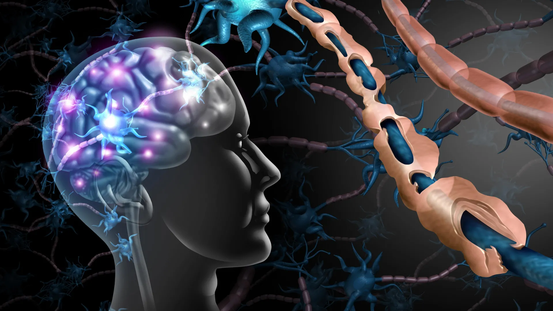
    Multiple sclerosis (MS) is a long-term autoimmune condition that affects over 2.9 million people around the world. In MS, the immune system mistakenly attacks the myelin sheath, a protective layer that insulates nerve fibers. This damage interrupts communication between the brain and body, leading to symptoms such as numbness, tingling, vision problems, and paralysis.

    Although existing treatments can help reduce inflammation, there are still no approved therapies that protect neurons or rebuild the damaged myelin sheath. Scientists have now made significant progress toward that goal with support from the National Multiple Sclerosis Society. Their work has led to the discovery of two compounds capable of promoting remyelination, the process of repairing the myelin coating on nerve fibers.

    The study, published in Scientific Reports, was led by Seema Tiwari-Woodruff, a professor of biomedical sciences at the University of California, Riverside, School of Medicine, and John Katzenellenbogen, a professor of chemistry at the University of Illinois Urbana-Champaign (UIUC). The research was funded through two National MS Society initiatives: a standard investigator-initiated grant and the organization’s Fast Forward program, which accelerates commercialization of promising research.

    “Our work represents more than a decade of collaboration, with the last four years focused on identifying and optimizing new drug candidates that show strong potential to treat MS and possibly other neurological diseases involving demyelination,” Tiwari-Woodruff said.

    With this support, the team launched a drug development program that has since been licensed by Cadenza Bio, Inc. Backed by investor funding, the company has continued advancing the research and is preparing for clinical testing of what could become a first-of-its-kind treatment for people with MS.

    From discovery to development

    This new work builds on earlier studies involving a compound called indazole chloride, which had shown promise in promoting myelin repair and regulating immune responses in mouse models of MS. However, indazole chloride lacked the chemical properties and patent potential required for clinical and commercial use, Tiwari-Woodruff explained.

    Working with UIUC chemists Katzenellenbogen and Sung Hoon Kim, who created new versions of the molecule, Tiwari-Woodruff’s group, led by recent UC Riverside graduate Micah Feri, screened more than 60 analogs of indazole chloride. From this effort, they identified two standout candidates, K102 and K110. Both showed better safety, efficacy, and drug-like characteristics in tests using mice and human cells.

    Among the two, K102 emerged as the leading candidate. It not only stimulated myelin repair but also helped regulate immune activity, a critical balance for MS therapies. The compound also performed well in human oligodendrocytes — cells responsible for producing myelin — derived from induced pluripotent stem cells, suggesting the results could translate effectively from animal studies to human disease.

    Normally, oligodendrocyte precursor cells develop into mature myelin-producing cells that repair nerve insulation. In MS, this repair process often breaks down, leading to lasting nerve damage. A compound like K102 that can restore myelin could help improve nerve signal transmission and potentially limit long-term disability.

    “K110 is also a strong candidate,” Tiwari-Woodruff said. “It has slightly different central nervous system effects and may be better suited for other conditions like spinal cord injury or traumatic brain injury, so we’re keeping it in the pipeline.”

    From bench to biotech

    Tiwari-Woodruff and Katzenellenbogen credit the National MS Society’s Fast Forward program as a turning point. Fast Forward accelerates the commercialization of promising therapies by promoting academic-industry partnerships. The highly competitive grant enabled Tiwari-Woodruff and Katzenellenbogen to generate sufficient data to license the rights to Cadenza Bio to develop K102 and K110. The patents are jointly held by UCR and UIUC, with an exclusive, worldwide licensing agreement in place between the universities and Cadenza Bio.

    “This project has been a good example of how long-standing academic collaborations can lead to real-world applications,” Katzenellenbogen said. “Our shared goal was always to take a promising idea and develop it into a therapy that could help people with MS. We’re finally getting close to that reality.”

    Initially, UCR’s Office of Technology Partnerships collaborated with UIUC to seek patent protection. Grace Yee, assistant director of technology commercialization at UCR, said the joint efforts of UCR, UIUC, and the National MS Society advocated for and promoted the technology to investors and industry for commercial development.

    “Our entrepreneurs-in-residence also helped advise the project, so the team was able to develop materials and messaging to highlight the project’s commercial value,” she said. “When investors expressed interest in the technology, UCR and UIUC helped them understand how the technology addresses an unmet need in treating MS. These efforts led to the licensing agreement with Cadenza Bio.”

    Elaine Hamm, chief operating officer at Cadenza Bio, said she and Carol Curtis, cofounder of Cadenza Bio, were impressed by the possibility of moving from slowing axon damage to repairing axon damage.

    “This is the future we want to build,” Hamm said. “It is why we licensed the technology, and why we are excited to move it forward to patients in need.”

    More than a decade in the making

    Tiwari-Woodruff and Katzenellenbogen have worked together for more than 12 years. Tiwari-Woodruff’s move from UCLA to UCR in 2014, she said, turned out to be a pivotal decision.

    “The support from UCR — from leadership to infrastructure — has been extraordinary,” Tiwari-Woodruff said. “None of this would’ve been possible without that backing. Funding for academic labs like mine and John’s is crucial. This is selfless work, driven by a deep love of science and commitment to human health.”

    Though the initial focus is MS, the team believes K102 and K110 could eventually be applied to other diseases involving neuronal damage, including stroke and neurodegeneration.

    Cadenza Bio is now advancing K102 through the necessary non-clinical studies required to support first-in-human clinical trials.

    “We’re hopeful that clinical trials can begin soon,” said Tiwari-Woodruff. “It’s been a long journey — but this is what translational science is all about: turning discovery into real-world impact.”

    The research was also supported in part by grants from the National Institutes of Health and Cadenza Bio.

    Tiwari-Woodruff, Katzenellenbogen, Kim, and Feri were joined in the research by Flavio D. Cardenas, Alyssa M. Anderson, Brandon T. Poole, Devang Deshpande, Shane Desfor, Kelley C. Atkinson, Stephanie R. Peterson, Moyinoluwa T. Ajayi, Fernando Beltran, Julio Tapia, and Martin I. Garcia-Castro of UCR; Kendall W. Nettles and Jerome C. Nwachukwu of The Scripps Research Institute, Florida; and David E. Martin and Curtis of Cadenza Bio, Oklahoma.

  • Study of the hippocampus suggests newborn brains start densely wired, then prune connections for sharper memory

    Study of the hippocampus suggests newborn brains start densely wired, then prune connections for sharper memory

    The hippocampus, a brain region essential for forming memories and mapping space, may develop in a way that challenges the long-held idea of the mind as a blank slate. New research from the Institute of Science and Technology Austria suggests key memory circuits begin life with unusually dense wiring that is later trimmed and refined.

    In a study published in Nature Communications, scientists examined how a major hippocampal network changes after birth in mice. The team focused on CA3 pyramidal neurons, cells widely seen as central to storing and retrieving memories.

    How the CA3 circuit develops

    Using patch-clamp recordings and high-resolution imaging, researchers compared the CA3 network across early postnatal stages, adolescence, and adulthood. The methods allowed them to measure tiny electrical signals and map how strongly neurons were connected at different ages.

    The results pointed to an early-life circuit that is highly connected and seemingly random, followed by a gradual shift toward fewer but more organized links. Rather than adding connections over time, the network became more efficient by losing many of its initial ones.

    Pruning may boost memory efficiency

    Lead researcher Peter Jonas said the pattern fits a pruning model in which the system starts full and then becomes streamlined. The researchers argue that an initially exuberant network could help the hippocampus quickly integrate different kinds of sensory information into usable memories.

    If the brain started with far fewer built-in links, the team notes, neurons would first need more time to find and connect to each other, potentially slowing early information processing. The study adds to broader evidence that brain development often involves overproduction of connections followed by activity-dependent pruning.

    While the work was conducted in mice, the hippocampus is highly conserved across mammals, making the findings relevant to ongoing debates about how genetics and experience shape learning. The authors say future research will need to clarify what signals drive which connections are kept or removed, and how this process relates to memory performance.

  • Ancient Lead Exposure and a Key Gene: New Clues to Why Modern Humans Outpaced Neanderthals

    Ancient Lead Exposure and a Key Gene: New Clues to Why Modern Humans Outpaced Neanderthals

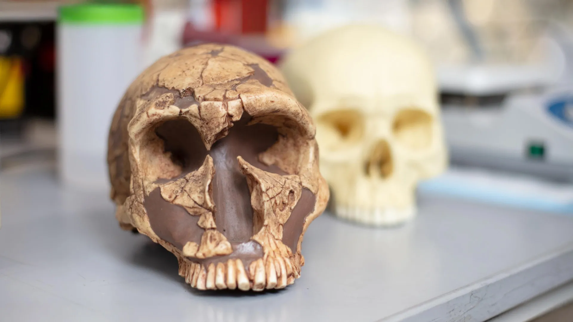
    What made the modern human brain so different from that of our extinct relatives, such as Neanderthals? Researchers at the University of California San Diego School of Medicine, along with an international team, have discovered that ancient hominids, including early humans and great apes, came into contact with lead far earlier than previously believed — up to two million years before modern humans began mining it. This long-term exposure may have influenced how early brains evolved, possibly hindering language and social development in all but modern humans, who possess a unique protective genetic variant. The findings were published in Science Advances on October 15, 2025.

    The team examined fossilized teeth from 51 hominids found across Africa, Asia, and Europe. The samples included both modern and archaic humans such as Neanderthals, early human ancestors like Australopithecus africanus, and extinct great apes including Gigantopithecus blacki.

    Lead traces were present in 73% of the fossils studied, with 71% of modern and archaic human samples showing contamination. Fossils of G. blacki dating back 1.8 million years revealed the highest levels of acute exposure.

    It was previously thought that humans began facing significant lead exposure only in recorded history, especially during the Roman era, when lead pipes were used for water systems, and later during the Industrial Revolution. Lead pollution declined only after the late twentieth century.

    “We stopped using lead in our daily lives when we realized how toxic it is, but nobody had ever studied lead in prehistory,” said corresponding author Alysson Muotri, Ph.D., professor of pediatrics and cellular & molecular medicine at UC San Diego School of Medicine, associate director of the Archealization Center, and director of the Sanford Integrated Space Stem Cell Orbital Research Center.

    To the researchers’ surprise, teeth from people born in the mid-twentieth century (the 1940s through the 1970s), when exposure to leaded gasoline and paint was widespread, showed similar lead patterns to ancient human fossils.

    The scientists suggest that ancient humans and their relatives might have encountered lead through their search for water, much like the Romans did later in history.

    “One possibility is that they were looking for caves with running water inside,” Muotri said. “Caves contain lead, so they were all contaminated. Based on the tooth enamel studies, it started very early in infancy.”

    Lead exposure disrupts brain growth and function, impairing intelligence and emotional regulation.

    Faced with this evidence, Muotri and his team began to question how modern humans managed to thrive despite such toxic conditions during their evolutionary past.

    A tiny genetic change

    A gene known as neuro-oncological ventral antigen 1 (NOVA1) plays a major role in brain formation and synaptic development. Acting as a key regulator of neurodevelopment, NOVA1 helps determine how neural progenitor cells react to lead exposure, and disturbances in its activity are linked to neurological disorders.

    Nearly all modern humans carry a version of the NOVA1 gene that differs by a single DNA base pair from the version found in Neanderthals. Earlier work from Muotri’s group showed that swapping the modern NOVA1 with the older variant in miniature brain models, called organoids, caused dramatic changes in brain structure and connectivity.

    “Everything about the organoids is identical except for that genetic variant, allowing us to ask whether that specific mutation between us and Neanderthals is giving us any advantage,” said Muotri. The archaic variant accelerated brain maturation but resulted in less complexity over time. “If all humans have this newer mutation in all corners of the world, very strong genetic pressure must have selected for it in our species.”

    To test whether lead exposure might have shaped this genetic shift, the researchers created brain organoids with both the modern and ancestral NOVA1 variants, exposing them to lead and monitoring the growth of cortical and thalamic neurons.

    They found that lead changed NOVA1 activity in both types of organoids, influencing genes linked to conditions such as autism and epilepsy.

    However, only the archaic NOVA1 variant altered the activity of FOXP2, a gene crucial for speech and language. People with certain FOXP2 mutations struggle to form complex words and sentences.

    “These type of neurons related to complex language are susceptible to death in the archaic version of NOVA1,” said Muotri. “ The FOXP2 gene is identical between us and the Neanderthals, but it’s how the gene is regulated by NOVA1 that likely contributes to language differences.”

    Evolutionary implications

    The findings suggest that the acquisition of the modern NOVA1 variant may have protected us from the detrimental effects of lead, promoting complex language development and social cohesion. This could have given modern humans a significant evolutionary advantage over Neanderthals, even in the presence of lead contamination.

    Muotri believes these results have important implications for understanding how environmental stressors shaped brain development during human evolution. He speculates that lead exposure may have contributed to the extinction of Neanderthals around 40,000 years ago.

    “Language is such an important advantage, it’s transformational, it is our superpower,” said Muotri. “Because we have language, we are able to organize society and exchange ideas, allowing us to coordinate large movements. There is no evidence that Neanderthals could do that. They might have had abstract thinking, but they could not translate that to each other. And maybe the reason is because they never had a system to communicate that was as efficient as our complex language.”

    Understanding how NOVA1 gene variants can affect FOXP2 expression helps elucidate the relationship between lead contamination and brain development and also sheds light on neurological conditions related to language, including speech apraxia — a condition that makes it difficult to produce speech sounds correctly — and autism.

    The study’s co-authors included Janaina Sena de Souza, Sandra M. Sanchez-Sanchez, Jose Oviedo, University of California San Diego; Marian Bailey and Matthew Tonge at Southern Cross University; Renaud Joannes-Boyau, Southern Cross University and University of Johannesburg; Justin W. Adams, University of Johannesburg and Monash University; Christine Austin, Manish Arora, Icahn School of Medicine at Mount Sinai, Kira Westaway, Macquarie University; Ian Moffat, Flinders University and University of Cambridge; Wei Wang and Wei Liao, Anthropology Museum of Guangxi; Yingqi Zhang, Institute of Vertebrate Paleontology and Paleoanthropology; Luca Fiorenza, Monash University and Johann Wolfgang Goethe University; Marie-Helene Moncel, Museum National d’Histoire Naturelle; Gary T. Schwartz, Arizona State University; Luiz Pedro Petroski and Roberto H. Herai, Pontifícia Universidade Católica do Paraná; Jose Oviedo, University of Arizona; and Bernardo Lemos, Harvard T. H. Chan School of Public Health.

    The study was funded, in part, by the National Institutes of Health (grants R01 ES027981, P30ES023515, R01ES026033), the Australian Research Council (grant DP170101597), the National Science Foundation (grant BCS 0962564), and the The Leakey Foundation.

    Disclosures: Muotri is the co-founder of and has an equity interest in TISMOO, a company specializing in genetic analysis and human brain organogenesis. The terms of this arrangement have been reviewed and approved by the University of California San Diego in accordance with its conflict-of-interest policies.

  • Ozempic and other GLP-1 drugs tied to lower depression and anxiety in large Swedish study, raising new questions about mental health benefits

    Ozempic and other GLP-1 drugs tied to lower depression and anxiety in large Swedish study, raising new questions about mental health benefits

    Popular GLP-1 medicines used for type 2 diabetes and weight loss, including Ozempic and Wegovy, may be linked to improved mental health outcomes, according to a large new analysis of Swedish health data. Researchers tracked changes in psychiatric care and work absence during periods when patients were using these drugs.

    The study, led by scientists from the University of Eastern Finland, Karolinska Institutet and Griffith University, examined national register data spanning 2009 to 2022. Nearly 100 000 people were included, with more than 20 000 having used a GLP-1 receptor agonist at some point during follow-up.

    What the study found

    Across the dataset, GLP-1 use was associated with fewer psychiatric hospital visits and fewer days of sickness absence related to mental health conditions. The strongest associations were reported for semaglutide, the active ingredient in Ozempic and Wegovy.

    During periods of semaglutide use, psychiatric hospital care and sickness absence were reported as 42% lower than during periods off treatment. The analysis also linked semaglutide exposure with a 44% lower risk of depression and a 38% lower risk of anxiety disorders.

    Signals beyond mood and anxiety

    The researchers also reported a lower rate of hospital care and work absence tied to substance use disorders during semaglutide treatment periods, with a reduction of 47%. They additionally found an association between GLP-1 receptor agonist use and reduced suicidal behavior, though the study design cannot determine causality.

    One author, Professor Mark Taylor of Griffith University, said the direction of the results aligned with earlier register-based research suggesting GLP-1 use may be associated with a reduced risk of alcohol use disorder. Because alcohol problems can worsen mood symptoms, the team said this could be one pathway behind the observed patterns.

    Why it may happen, and what it means

    Experts caution that registry studies can show links but cannot prove the drugs directly prevent depression or anxiety, since unmeasured factors could influence who starts or stays on treatment. Researchers said potential explanations range from improved blood sugar control and weight loss to reduced alcohol use and better quality of life.

    They also pointed to possible direct effects on the brain, including changes in reward-related pathways, as a hypothesis that needs targeted clinical research. The findings were published in The Lancet Psychiatry, adding weight to an ongoing debate as regulators and clinicians continue to watch for both potential benefits and risks affecting mental health.