Blog

  • Penn State-linked study suggests perceived control can cut daily stress by driving faster problem-solving

    Penn State-linked study suggests perceived control can cut daily stress by driving faster problem-solving

    New research involving scientists affiliated with Penn State suggests that feeling more in control of everyday problems can make people more likely to resolve them, easing day-to-day stress. The findings add to evidence that small psychological shifts can influence how people respond to routine hassles.

    The study, published in Communications Psychology, analyzed daily reports from more than 1 700 adults participating in the National Study of Daily Experiences, a project linked to the broader MIDUS health survey. Participants logged stressors over eight consecutive days and noted whether each issue was resolved by day’s end.

    Control and stress resolution link

    Researchers found that on days when individuals reported greater perceived control over a stressor, they were substantially more likely to take action that led to resolution. Examples included addressing an interpersonal conflict, handling a home problem, or responding to work overload.

    The analysis indicated that perceived control varies from day to day rather than functioning as a fixed personality trait. That matters because it suggests control can potentially be strengthened through context, planning, or support, rather than being something people either have or lack.

    Why age may amplify effects

    The link between perceived control and resolving stressors appeared to strengthen with age across the two survey waves taken roughly a decade apart. Researchers reported that later in the study period, higher-than-usual perceived control was associated with an even greater likelihood of resolving the day’s stressor.

    Scientists cautioned that perceived control does not remove stressors, but it may help people respond in ways that prevent problems from lingering. The authors say the next step is to examine whether faster resolution could also reduce the health impact of chronic stress over longer periods.

  • A simple blood test could spot depression risk early by tracking immune cell aging

    A simple blood test could spot depression risk early by tracking immune cell aging

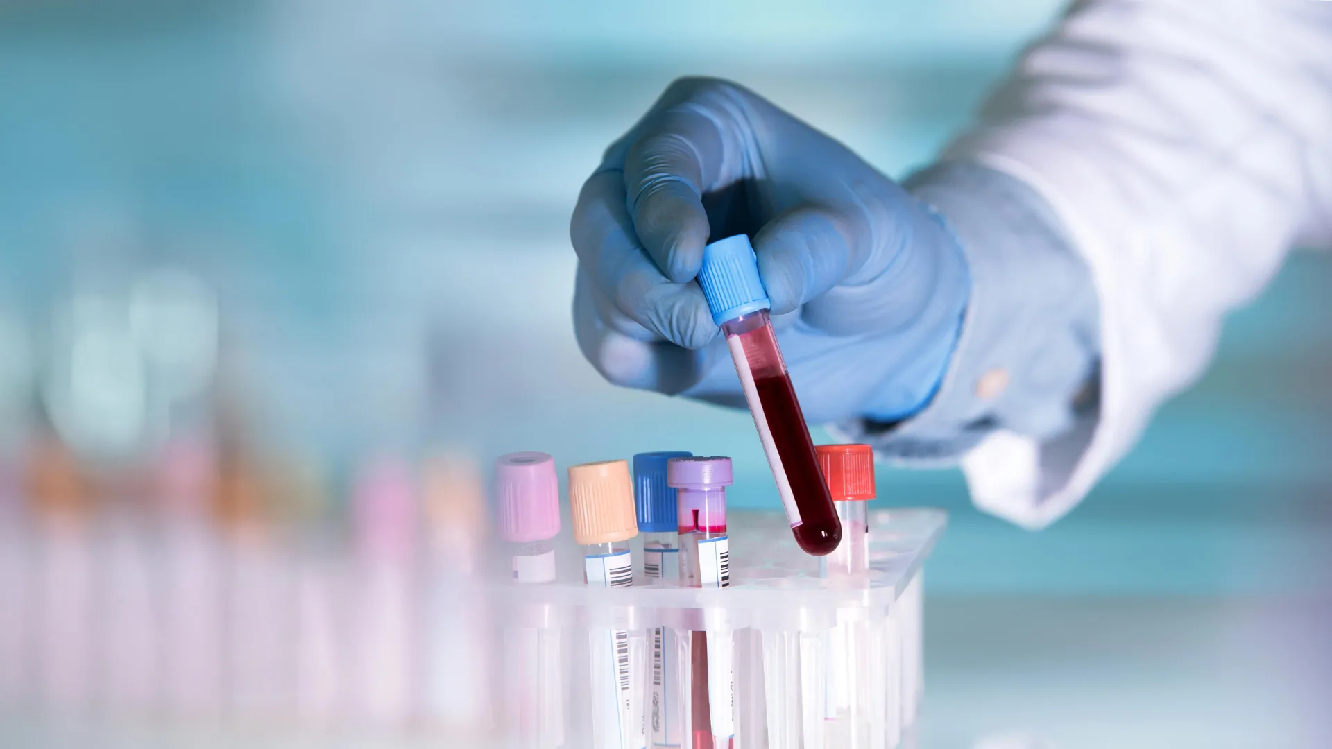
    Researchers are testing whether a routine blood sample could help flag depression earlier, using measures of biological aging in specific immune cells. The work adds to a growing push for objective biomarkers that could complement symptom-based mental health screening.

    The study, published in The Journals of Gerontology: Series A, examined epigenetic changes that act like molecular timestamps on DNA. These so-called epigenetic clocks estimate biological age, which can advance faster than chronological age under stress, illness, or other factors.

    Why depression has been hard to test

    Depression is typically diagnosed through clinical interviews and questionnaires, not lab confirmation, partly because the condition can look very different across patients. Some people primarily experience somatic symptoms such as sleep and appetite changes, while others struggle most with mood and cognition, including hopelessness and loss of pleasure.

    That variability can complicate detection, especially when physical symptoms overlap with other chronic conditions. Clinicians may order bloodwork to rule out medical causes, but there is still no widely accepted biological test that can confirm depression on its own.

    Monocytes emerge as a key signal

    The research analyzed data from 440 women, including 261 living with HIV and 179 without HIV, drawing on the long-running Women’s Interagency HIV Study. Depression symptoms were assessed using the Center for Epidemiologic Studies Depression Scale, a 20-item tool that captures both somatic and non-somatic features.

    Blood samples were used to calculate epigenetic aging in two ways: a broad measure across multiple cell types and a monocyte-focused clock. Monocytes are white blood cells involved in immune responses and inflammation, processes increasingly studied for their links to mental health.

    The monocyte-specific aging measure tracked most strongly with non-somatic depression symptoms such as anhedonia, hopelessness, and a sense of failure, in women with and without HIV. By contrast, the broader multi-tissue aging measure did not show the same relationship, suggesting the signal may be cell-type specific.

    What this could change in care

    The authors caution that the findings are not yet ready for clinical use and do not mean a single blood draw can diagnose depression today. Larger studies, replication in different populations, and clearer thresholds would be needed before a test could be validated for real-world screening.

    Still, the results point to a possible path toward earlier, more precise detection, particularly for people whose physical symptoms might be attributed to other illnesses. If confirmed, immune-cell epigenetic measures could also support more personalized care by helping researchers distinguish depression subtypes and refine treatment matching.

  • How reward bias can make lies feel true, especially from friends

    How reward bias can make lies feel true, especially from friends

    New neuroscience research is adding detail to a familiar problem: people often struggle to spot dishonesty, particularly when the message sounds beneficial. The findings suggest that the promise of a gain can subtly weaken how carefully we evaluate whether information is true.

    The study, led by Yingjie Liu of North China University of Science and Technology, tested how people judge messages depending on who delivers them. Researchers focused on whether trust shifts when information comes from a friend versus a less familiar person.

    Inside the brain during deception

    Using brain imaging with 66 healthy adults, the team examined neural activity while participants exchanged information through computer screens. Messages were framed around outcomes described as gains or losses, allowing scientists to track how reward and risk contexts shape belief.

    Across the experiment, participants were more likely to accept false information in gain situations. Brain regions linked to reward processing, risk assessment and interpreting others’ intentions showed patterns consistent with relaxed scrutiny when a positive outcome seemed possible.

    Why friends can be persuasive

    Friendship added another layer: when a friend delivered the potentially misleading message, the pair showed synchronized brain activity. That alignment shifted with context, strengthening in reward-related areas during gains and in risk-related areas during losses.

    Researchers reported that these shared neural patterns helped predict when someone was most likely to be misled by a friend. The results point to a mechanism in which social closeness and reward expectations combine to make certain claims feel credible even when they should prompt doubt.

    While the work does not mean people always trust friends blindly, it highlights a consistent vulnerability. In everyday decisions, offers that appear mutually beneficial may deserve extra verification, precisely because the brain can treat them as safer than they are.

  • Creatine research shifts beyond the gym: What studies suggest about brain health, mood and aging

    Creatine research shifts beyond the gym: What studies suggest about brain health, mood and aging

    Creatine has long been associated with strength and sprint performance, but a growing body of research is examining what it may do beyond muscle gains. Scientists are increasingly focused on its role in cellular energy, including in the brain, where energy demand is high.

    The compound is made in the body from amino acids and stored mainly in skeletal muscle, with smaller amounts in organs such as the brain and heart. In cells it supports the rapid recycling of ATP, the core energy molecule, which helps explain why supplementation can aid short, high-intensity efforts.

    Why creatine matters in the brain

    Because brain tissue depends on steady energy supply, researchers are studying whether creatine can support cognition under stress, sleep loss or aging. Some studies have reported improvements in tasks linked to memory and processing speed, especially in people likely to start with lower creatine stores.

    Interest is also expanding into mental health and neurological disease, including depression and Parkinson’s disease, where energy metabolism and inflammation may play a role. Evidence remains mixed and condition-specific, and experts stress that early signals do not yet translate into routine medical use.

    What dosing research typically uses

    The most studied form is creatine monohydrate, commonly taken either as a short loading phase of about 20 g daily for 5 to 7 days or as a steady daily dose around 3 to 5 g. Research suggests both approaches can raise muscle stores, with loading working faster and lower daily dosing reaching saturation over several weeks.

    Absorption and retention vary between individuals, partly because tissues have a storage ceiling and excess is converted to creatinine and excreted. Some evidence indicates taking creatine with carbohydrates can increase uptake, though the real-world impact differs by person and diet.

    Safety, limits and who should ask first

    Large reviews and position statements have generally found creatine to be safe for healthy adults when used as studied, and it is not a steroid or hormone. The most common issues are practical rather than dangerous, such as water retention or stomach upset at higher single doses.

    Clinicians still advise caution for people with kidney disease or those advised to limit protein-related metabolites, since creatinine levels can rise with supplementation and complicate lab interpretation. Researchers emphasize that creatine is not a cure-all and works best as a targeted aid alongside training, nutrition and medical guidance when needed.

  • Deep Sleep and Growth Hormone: UC Berkeley Study Maps Brain Circuit That Links Nighttime Repair to Wakefulness

    Deep Sleep and Growth Hormone: UC Berkeley Study Maps Brain Circuit That Links Nighttime Repair to Wakefulness

    Deep non-REM sleep has long been tied to the body’s biggest overnight growth hormone surge, a rhythm linked to tissue repair, muscle maintenance and metabolic health. A new study from researchers at the University of California, Berkeley offers a clearer explanation of how the brain coordinates that hormone release during sleep.

    In work published in Cell, the team traced a neural circuit in the hypothalamus that helps control when growth hormone is released and how that signal is kept in balance. The findings come as sleep disruption is increasingly associated in research with higher risks for weight gain, insulin resistance and cardiovascular disease.

    A circuit with two hormone controls

    The study focuses on two hypothalamic signals that act as opposing levers: growth hormone releasing hormone, which promotes growth hormone output, and somatostatin, which suppresses it. By mapping how these systems behave across sleep stages, researchers aimed to explain why fragmented or reduced deep sleep can blunt growth hormone release.

    Using mice, the researchers recorded neural activity and manipulated specific neurons to observe how these signals shift across REM and non-REM sleep. They reported that the two hormones follow distinct patterns depending on the sleep stage, producing different growth hormone dynamics over the night.

    How growth hormone feeds back

    The team also describes a feedback loop connecting growth hormone signaling to the locus coeruleus, a brainstem hub involved in alertness and attention. As growth hormone levels build, the circuit can influence arousal, suggesting the hormone is not only an output of sleep but also a contributor to sleep-wake regulation.

    Because the locus coeruleus is implicated in a range of neurological and psychiatric conditions, the authors say the circuit-level map could help guide future work on sleep disorders and diseases where arousal systems are disrupted. However, the research was conducted in mice, and any clinical applications would require further validation in humans.

    Why the findings matter

    Growth hormone is best known for its role in childhood and adolescent growth, but it also supports adult physiology by influencing body composition and how the body handles sugar and fat. That is why chronically poor sleep, particularly reduced deep sleep, has been linked in broader research to metabolic problems over time.

    The researchers argue that identifying the wiring behind growth hormone regulation may eventually inform therapies that target specific nodes in the sleep-hormone system. For now, the study adds detailed biological context to a familiar health message: sleep quality can shape key hormonal processes, not just next-day energy.

    The work was supported by the Howard Hughes Medical Institute and the Pivotal Life Sciences Chancellor’s Chair fund, and included collaborators from UC Berkeley and Stanford University. The authors emphasize that the new circuit map is a foundation for future studies on how sleep architecture and hormones interact in health and disease.

  • A lasting brain switch in addiction and stress: Why ΔFosB is drawing new attention in mental health research

    A lasting brain switch in addiction and stress: Why ΔFosB is drawing new attention in mental health research

    Advances in molecular psychiatry are sharpening scientists’ view of how stress and drugs can leave long-lasting marks on the brain. A recent interview published by Genomic Press in the journal Brain Medicine highlights decades of work that helped connect fleeting experiences to persistent changes in behavior.

    In the discussion, neuroscientist Eric J. Nestler, dean of the Icahn School of Medicine at Mount Sinai, traces how early training in brain chemistry led him to focus on the biology behind addiction, depression and resilience. He describes a field that has moved from broad theories toward specific molecules, cell types and circuits that can be measured.

    ΔFosB and long-term brain change

    One of the best-known findings from this line of research centers on ΔFosB, a transcription factor that can build up in reward-related brain circuits after repeated drug exposure and prolonged stress. Unlike many proteins that degrade quickly, ΔFosB can persist for weeks, helping to explain how short periods of exposure may trigger longer-lasting shifts in gene activity.

    Researchers have linked this durability to changes in motivation and reward processing that can raise vulnerability to addiction. The idea is not that one factor explains complex disorders, but that stable molecular signals like ΔFosB can act as a biological bridge between experience and enduring neural adaptation.

    From epigenetics to single-cell tools

    Nestler also points to a major methodological shift in the field, from studying signaling pathways to mapping gene regulation through epigenetic mechanisms such as chromatin modifications. Those approaches have been accelerated by tools that can parse differences across brain regions and, increasingly, across individual neuron types.

    Single-cell methods are now enabling researchers to look for patterns that may be missed when tissue is analyzed in bulk. That trajectory is feeding interest in whether future treatments could be tailored more precisely to particular circuits or cell populations involved in mood and substance-use disorders.

    Why resilience is becoming central

    A notable theme in the interview is a push to study resilience, not only pathology. Experiments in animals have identified molecular and circuit signatures associated with maintaining normal behavior despite stress, raising the possibility of therapies designed to strengthen protective mechanisms.

    Some resilience-oriented strategies are already being tested clinically for depression, reflecting a broader shift toward interventions that aim to improve adaptive capacity as well as relieve symptoms. The interview argues that focusing on what helps certain individuals recover could open complementary pathways for drug development.

    Nestler also underscores the importance of linking animal findings with human evidence, including results from postmortem brain studies in people affected by addiction and stress-related conditions. He warns that politicizing science could slow progress, stressing that medical research should remain independent and broadly beneficial.

  • Astrocytes move into the spotlight: New Nature study links overlooked brain cells to fear memories and PTSD pathways

    Astrocytes move into the spotlight: New Nature study links overlooked brain cells to fear memories and PTSD pathways

    Brain research is increasingly challenging the long-held idea that neurons alone drive fear and trauma responses. A new study in Nature points to astrocytes, star-shaped support cells, as active players in how fear memories are formed, recalled and reduced.

    Astrocytes are widely distributed throughout the brain and have traditionally been seen as caretakers that keep neural circuits stable. The new work suggests they can also shape signaling in the amygdala, a central hub for processing threat and generating fear-related learning.

    What the researchers observed

    Using a mouse model of fear learning, scientists tracked astrocyte activity in real time with fluorescent sensors. Astrocyte signaling rose during fear conditioning and again during memory recall, then declined as fear responses weakened through extinction training.

    The team also manipulated how astrocytes communicate with nearby neurons. Enhancing astrocyte-to-neuron signaling strengthened fear expression, while dampening those signals reduced fear responses, indicating astrocytes can tune the intensity of fear memories.

    How it changes the fear circuit

    When astrocyte activity was disrupted, neurons in the amygdala had difficulty forming the typical activity patterns associated with fear. That interference also appeared to affect how defensive-response information is routed to other brain regions involved in choosing and executing behavior.

    Researchers reported effects beyond the amygdala, including changes in fear-related signaling reaching the prefrontal cortex, an area tied to decision-making and regulation of emotional responses. The results suggest astrocytes may influence how the brain decides whether a threat response is appropriate.

    Why it matters for PTSD

    PTSD and several anxiety disorders are marked by persistent, hard-to-extinguish fear memories and heightened reactions to cues that are no longer dangerous. If astrocytes help govern both the expression and the fading of fear, they could become a complementary target alongside neuron-focused approaches.

    The researchers caution that translating mouse findings to human treatments takes time, but the study reframes fear circuitry as a partnership between neurons and glia. Next steps include mapping astrocyte roles across the wider threat network, including regions that coordinate freezing and flight responses.

  • Warm hugs and the brain: New thermoception research reveals how temperature shapes body awareness and mood

    Warm hugs and the brain: New thermoception research reveals how temperature shapes body awareness and mood

    New neuroscience research is sharpening the picture of why a warm hug can feel uniquely calming, suggesting that temperature signals from the skin help the brain maintain a stable sense of the body. A recent review argues that thermoception, the ability to sense warmth and cold, plays a larger role in emotion and self-awareness than previously assumed.

    Published in Trends in Cognitive Sciences, the review brings together evidence from psychology, neuroscience and clinical studies to show that temperature is not just about comfort or survival. Instead, thermal cues appear to influence how strongly people experience their body as their own, a process often described as body ownership.

    Temperature as a body-brain signal

    Researchers highlight that thermoception works alongside touch and internal bodily signals to shape moment-to-moment awareness of the self. Warmth in particular is framed as a biologically meaningful cue of safety and care, learned early in life and reinforced through social contact.

    Laboratory work has linked warm, gentle contact to neural pathways that feed into brain regions involved in interoception, including the insular cortex. These circuits help integrate what the body feels from the outside with internal state, supporting emotional regulation during close social interactions.

    Links to mental health conditions

    The review points to clinical observations in which disrupted body awareness is common, including depression, anxiety, trauma-related disorders and eating disorders. In these settings, people may describe feeling detached from their body or less certain about bodily sensations.

    Studies in conditions such as stroke, anorexia nervosa and body integrity dysphoria suggest that altered thermal perception can occur alongside disturbances in body ownership. The authors argue this overlap makes temperature-sensing a promising, if underused, lens for understanding symptoms that involve disconnection from the body.

    From therapy to prosthetics

    Beyond explaining everyday comfort, the authors suggest thermoception research could inform sensory-based approaches in rehabilitation and mental health care. Better mapping of skin-to-brain temperature pathways may help clinicians identify vulnerabilities and tailor interventions that work through controlled sensory input.

    Engineers could also apply these insights to prosthetics, where adding realistic thermal feedback may improve how naturally an artificial limb is experienced. The review further notes that more frequent exposure to extreme heat and cold could affect mood and stress, making temperature an emerging topic in public health research.

    In practical terms, the science helps explain why warm social touch can be grounding: thermal and tactile signals arrive together, reinforcing the brain’s model of the body in a context associated with safety. That combination may be one reason a brief, warm hug can feel like both physical comfort and emotional reassurance.

  • UCSF study flags FTL1 protein as a driver of brain aging, and a potential new target to restore memory

    UCSF study flags FTL1 protein as a driver of brain aging, and a potential new target to restore memory

    Aging can take a heavy toll on the hippocampus, the brain region central to learning and memory. Researchers at the University of California, San Francisco report they have identified a protein that may play an outsized role in that decline.

    In a study published in Nature Aging, the team points to FTL1, a ferritin-related protein involved in iron handling, as a key molecular change seen in older mouse hippocampus. The researchers say shifting FTL1 levels altered memory performance and the strength of neural connections in ways that tracked with age.

    A standout signal in aging brains

    To pinpoint what changes over time, scientists compared gene and protein patterns in the hippocampus of young and older mice. Among many measurements, FTL1 emerged as the most consistent difference between age groups.

    Older mice had higher FTL1 levels alongside fewer synaptic connections and worse results on cognitive tasks. The authors report that the pattern suggested more than a passive marker of aging, raising the possibility that FTL1 helps drive the process.

    What happened when FTL1 was altered

    When researchers increased FTL1 in young mice, the animals developed brain and behavioral changes resembling those seen in older mice. The hippocampus showed reduced connectivity, and performance on memory-related testing declined.

    Cell experiments offered a potential explanation: neurons pushed to make more FTL1 formed simpler structures, with fewer branching extensions needed for complex signaling. That shift, the team argues, could help explain how elevated FTL1 weakens hippocampal circuitry.

    Can memory decline be reversed?

    In older mice, lowering FTL1 was linked to improved synaptic connections and better memory test performance. Senior author Saul Villeda said, “It is truly a reversal of impairments,” emphasizing that the effect went beyond delaying decline.

    The group also reported a metabolic component, with higher FTL1 associated with slower energy use in hippocampal cells. In lab settings, boosting cellular metabolism with an experimental compound reduced the harmful effects tied to elevated FTL1, pointing to possible therapeutic angles.

    Experts caution that mouse findings do not automatically translate to human brain aging, and any treatment approach would require extensive safety and efficacy testing. Still, the UCSF team argues that targeting FTL1 or related metabolic pathways could eventually open a new route for interventions aimed at age-related cognitive decline.

  • Japanese researchers grow thalamus-cortex brain circuits in lab assembloids, opening a new window on neural development

    Japanese researchers grow thalamus-cortex brain circuits in lab assembloids, opening a new window on neural development

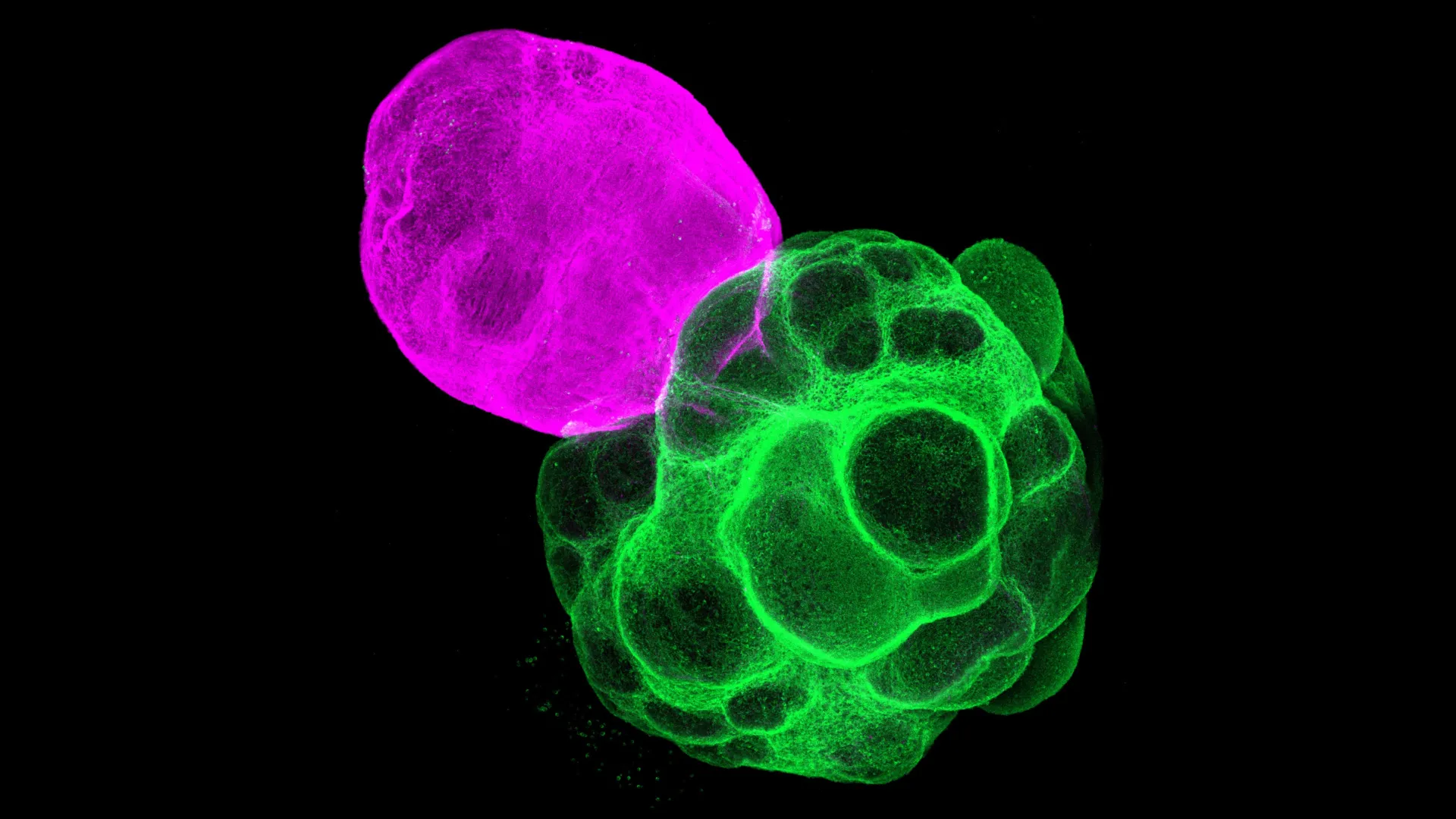
    Researchers in Japan have recreated key human brain circuits in the lab by fusing miniature models of the thalamus and cerebral cortex into what are known as assembloids. Built from human induced pluripotent stem cells, the multi-region tissue is designed to mimic how developing brain areas connect and coordinate activity.

    The work, published in the Proceedings of the National Academy of Sciences, focuses on the thalamus-cortex pathway that helps organize sensory processing and higher cognition. Because direct studies of early human brain wiring are limited by ethical and technical constraints, organoid-based models have become an important alternative.

    Why thalamus-cortex wiring matters

    The cerebral cortex relies on precisely timed communication among different neuron types and between distant brain regions. Disruptions in these networks are linked to neurodevelopmental conditions, including autism spectrum disorder, making circuit formation a high-priority target for basic and translational research.

    Animal studies have long suggested the thalamus helps shape cortical development, but confirming the details in human tissue has been difficult. The new assembloid model aims to capture those cross-region interactions more realistically than single-region organoids.

    How the assembloids were built

    The team first grew separate thalamic and cortical organoids from iPS cells and then fused them to allow fibers to extend between regions. Over time, axons from each side grew toward the other and formed synapses, resembling the bidirectional connectivity seen in the developing brain.

    When researchers compared cortical tissue grown alone with cortical tissue connected to the thalamus, the connected cortex showed gene activity consistent with greater maturation. The results support the idea that thalamic input can accelerate aspects of cortical development.

    Signals that synchronize specific neurons

    To probe function, the scientists tracked how activity traveled through the fused tissue and observed wave-like patterns moving from the thalamus into the cortex. That flow was associated with synchronized activity across parts of the cortical network.

    The effect differed across major cortical excitatory neuron classes, with synchronized patterns seen in pyramidal tract and corticothalamic neurons that communicate with the thalamus. Intratelencephalic neurons, which do not project to the thalamus, showed less synchrony, suggesting thalamic input selectively reinforces certain circuit types.

    Researchers say the platform could help clarify how human circuits assemble at the level of cell types and connections, and provide a testbed for studying disease mechanisms. In the longer term, thalamus-cortex assembloids may also support screening strategies aimed at restoring or stabilizing abnormal network activity.