Blog

  • Parkinson’s Study Points to SCAN Brain Network as a New Treatment Target, With Early Gains From Non-Invasive Stimulation

    Parkinson’s Study Points to SCAN Brain Network as a New Treatment Target, With Early Gains From Non-Invasive Stimulation

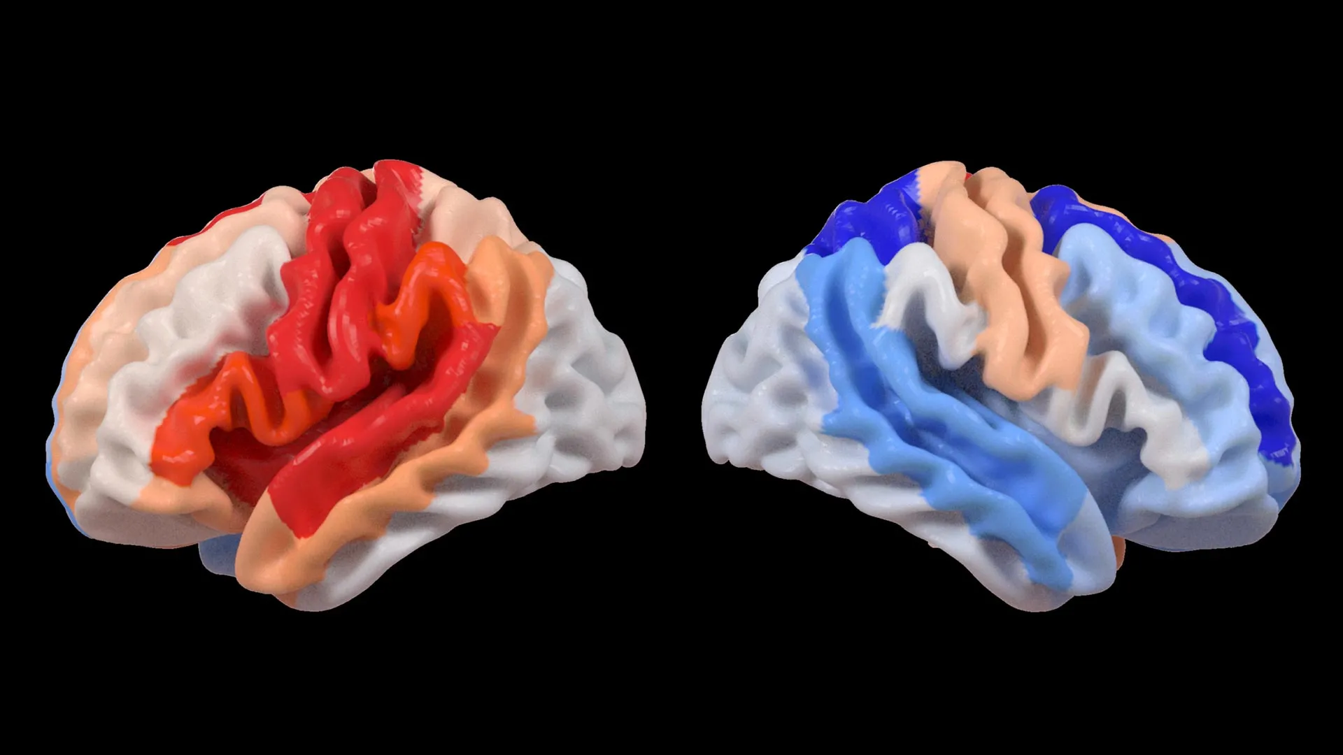
    Researchers say they have identified a specific brain network that may sit at the core of Parkinson’s disease, potentially reshaping how the condition is understood and treated. The findings focus on the somato-cognitive action network, or SCAN, which links planning and thinking with physical movement.

    Parkinson’s is a progressive neurological disorder affecting more than 10 million people worldwide, commonly causing tremor, stiffness, slowed movement, sleep disruption and cognitive changes. Standard therapies such as levodopa can ease symptoms for years, while deep brain stimulation can help selected patients, but neither stops disease progression.

    A network view of Parkinson’s

    The international team, led by China’s Changping Laboratory with collaborators including Washington University School of Medicine in St. Louis, analyzed brain imaging data from more than 800 participants across several centers. The dataset included people with Parkinson’s receiving different treatments, along with healthy volunteers and people with other movement disorders for comparison.

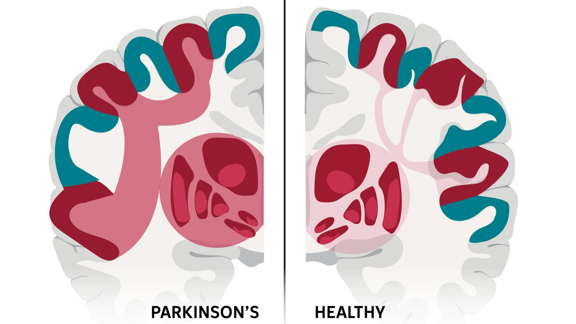
    The researchers report that Parkinson’s is marked by unusually strong connectivity between SCAN and deeper brain structures in the subcortex. Across multiple therapies examined, the study found that treatments tended to work better when they reduced this excessive coupling rather than simply stimulating nearby regions.

    Non-invasive stimulation shows early promise

    Building on that signal, the team tested a personalized, high-precision approach using transcranial magnetic stimulation, a non-invasive technique that delivers magnetic pulses through the scalp. In a small trial, 18 patients who received SCAN-targeted stimulation showed a higher response rate after two weeks than 18 patients who received stimulation near, but not on, the SCAN target.

    In the SCAN-targeted group, 56% met the study’s response threshold, compared with 22% in the comparison group, a roughly 2.5-fold difference. The work suggests that matching stimulation more precisely to an individual’s SCAN anatomy could improve outcomes, though larger and longer studies are needed.

    What comes next for SCAN targeting?

    The authors caution that the study does not prove SCAN changes cause Parkinson’s, and the non-invasive results are early-stage. They also note that more basic research is needed to map how different SCAN subregions relate to specific symptoms such as gait, tremor, mood and cognition.

    Even so, the findings add momentum to efforts to move neuromodulation earlier in the disease course, when symptoms are still developing and disability is lower. The researchers say future trials will explore additional non-invasive approaches, including surface electrode stimulation and low-intensity focused ultrasound, to influence SCAN activity more precisely.

    The study was published in Nature on Feb. 4 and was supported by funding from U.S. National Institutes of Health programs and major Chinese research grants. The authors also disclosed multiple industry relationships and patent interests related to neuromodulation and targeting tools, which were reported as managed under institutional conflict-of-interest policies.

  • Precision Treatment for Depression: A New Data-Driven Model Aims to Match Patients With the Therapy Most Likely to Work

    Researchers are moving beyond trial-and-error care for depression with a precision approach designed to better match patients to treatments based on individual characteristics. The effort reflects growing evidence that depression symptoms and recovery paths vary widely from person to person.

    The project, led by psychologists at the University of Arizona and Radboud University, draws on patient-level data from randomized clinical trials across the world. Their protocol, published in PLOS One, outlines how they plan to build a clinical decision support tool for adult depression treatment selection.

    Why first-line care often fails

    Standard care frequently begins with a first-line medication or therapy and then shifts if symptoms persist, a process that can take months. The researchers point to prior findings that roughly half of patients do not respond to an initial treatment, highlighting the need for better targeting.

    Instead of offering broad guidelines, the planned tool would generate a single recommendation by weighing multiple factors at once. These include demographic information such as age and gender, along with clinical features like anxiety symptoms or personality-related difficulties.

    What data the model will use

    The team aggregated outcomes from more than 60 clinical trials involving nearly 10 000 patients, covering several widely used interventions. The treatments include antidepressant medications and multiple psychotherapy approaches, such as cognitive therapy, behavioral therapy, interpersonal therapy and short-term psychodynamic therapy.

    By combining many trials, the researchers aim to overcome limits that can affect prediction models built from single studies with smaller samples. They say the work required years of data cleaning and harmonization before analysis could begin.

    When it could reach clinics

    The next step is to develop the algorithm and then test it in a clinical trial to see whether tool-guided care improves outcomes compared with usual practice. If the results hold up, the system could be deployed as a simple software or web-based application used during routine assessments.

    The researchers argue that the inputs are intentionally practical, relying on information that can be collected through standard questionnaires and basic clinical intake. Their longer-term goal is to help clinicians and patients reach effective treatment faster while using existing mental health resources more efficiently.

  • Late-life depression could precede Parkinson’s or Lewy body dementia, Danish study suggests

    Late-life depression could precede Parkinson’s or Lewy body dementia, Danish study suggests

    While there is currently no cure for Parkinson’s disease or Lewy body dementia, addressing depression early could improve quality of life and overall care for patients as these diseases develop.

    study published in General Psychiatry provides the most detailed longitudinal evidence to date, demonstrating that depression frequently precedes the diagnosis of PD and LBD and remains elevated for several years thereafter.


    Drawing on comprehensive Danish national health registers, the researchers conducted a retrospective case–control study including 17,711 individuals diagnosed with PD or LBD between 2007 and 2019. Researchers compared these patients with people of similar age and sex who were diagnosed with other long-term conditions, including rheumatoid arthritis, chronic kidney disease, and osteoporosis.


    The results showed a clear pattern: depression occurred more often and earlier in people who went on to develop Parkinson’s disease or Lewy body dementia than in those with other chronic illnesses. In the years leading up to diagnosis, the risk of depression rose steadily, peaking in the three years before diagnosis. Even after diagnosis, patients with Parkinson’s disease or Lewy body dementia continued to experience higher rates of depression than the comparison groups.


    Importantly, this pattern could not be fully explained by the emotional burden of living with a chronic illness. Other long-term diseases that also involve disability did not show the same strong increase in depression risk. This suggests that depression may be linked to early neurodegenerative changes in the brain, rather than being only a psychological reaction to declining health.


    The findings were especially striking for Lewy body dementia, where rates of depression were even higher than in Parkinson’s disease, both before and after diagnosis. Researchers note that differences in disease progression and brain chemistry may help explain this trend.


    “Following a diagnosis of PD or LBD, the persistent higher incidence of depression highlights the need for heightened clinical awareness and systematic screening for depressive symptoms in these patients.” first author Christopher Rohde noted “Thus, our main conclusion—that PD/LBD are associated with a marked excess depression risk preceding and following diagnosis when compared with other chronic conditions—remains valid.”


    The authors emphasize that this does not mean everyone with depression will develop Parkinson’s disease or dementia. Instead, they recommend greater awareness and closer monitoring when depression appears for the first time in older adults.


    While there is currently no cure for Parkinson’s disease or Lewy body dementia, addressing depression early could improve quality of life and overall care for patients as these diseases develop.

  • Study finds AI still struggles to read social cues in video, a hurdle for self-driving cars and robots

    Humans still outperform today’s artificial intelligence at interpreting social interactions in moving scenes, a skill that underpins safer self-driving cars and more helpful assistive robots. New research from Johns Hopkins University suggests many leading models miss context that people grasp quickly.

    The team examined how well AI systems can infer intentions, relationships, and ongoing actions when people share a scene. These judgments help determine whether two pedestrians are chatting, about to cross the street, or reacting to one another.

    Testing AI against human perception

    In the study, participants watched three-second video clips and rated social features on a one-to-five scale. The clips showed people interacting, doing side-by-side activities, or acting independently.

    Researchers then asked more than 350 AI language, video, and image models to predict human ratings and expected brain responses. For large language models, the systems evaluated short, human-written captions describing the videos.

    Where models fell behind

    People largely agreed with one another across questions, but the AI models did not show the same consistency, regardless of size or training data. Video models often struggled to describe what people were doing, and image models given still frames could not reliably detect communication.

    Language models were comparatively better at predicting how humans would judge behavior, while video models were more aligned with predicted neural activity. Even so, none of the model types matched human responses across the board.

    Why reading the room is hard

    The researchers argue the gap highlights a difference between recognizing objects in static images and understanding the unfolding story in real life. They suggest a potential cause is that many AI architectures draw inspiration from brain systems tuned for static vision rather than dynamic social scenes.

    Lead author Leyla Isik said an autonomous vehicle needs to read intentions and goals, not just identify people and objects. Co-first author Kathy Garcia added that social relationships, context, and dynamics appear to be a persistent blind spot in current model development.

    The findings are being presented at the International Conference on Learning Representations, where researchers will discuss implications for AI that must interact safely with humans. The work adds to a growing body of evidence that high scores on benchmarks do not always translate to robust real-world understanding.

  • Study Tracks Overimitation in Infants: Copying Starts Early, but In-Group Bias Comes Later

    Infants begin copying unnecessary actions well before age two, but that early tendency does not yet appear tied to choosing people who seem more similar to them. That is the central finding of a new Concordia University study published in Frontiers in Developmental Psychology.

    Overimitation refers to copying steps that are irrelevant to achieving a goal, such as repeating an extra action when opening a box to reach a toy. Researchers have long linked the behavior in older children to social affiliation, but evidence in children under two has been limited.

    What the new study tested

    The team observed 73 children aged 16 to 21 months, with an average age just over 18 months. Each child completed four tasks designed to measure different types of imitation and a separate test for in-group preference.

    In the overimitation task, an adult demonstrated three steps to open a box, including one action that did not help retrieve the toy. Other tasks measured memory-based copying and the ability to infer an adult’s intended goal when the adult appeared to fail at completing an action.

    Low overimitation, no in-group pull

    The researchers found low levels of overimitation at this age, and children’s performance was not driven by in-group preference. In the in-group task, children chose between objects offered by a woman and a robot, and their choices did not predict overimitation.

    By contrast, two other imitation measures showed a clear relationship: elicited imitation, often used to assess early memory, and imitation of unfulfilled intentions. This pattern suggests that at 16 to 21 months, imitation is more closely aligned with developing cognition and recall than with group-based social motivations.

    Why it matters for parents and educators

    The authors argue that the social reasons often proposed for overimitation may emerge later in development, as children learn more about group membership. They point to related work indicating that by around preschool age, overimitation can align more with preferences for similar peers.

    The findings also serve as a reminder that very young children may copy both helpful and unnecessary behaviors from adults. The researchers say this has implications for modeling actions in homes and classrooms where early learning is strongly shaped by observation.

  • MIT’s ComMAND gene circuit could make gene therapy dosing safer and more predictable

    Gene therapy has long promised one-time treatments for disorders caused by a missing or faulty gene, but controlling how strongly a delivered gene turns on in cells remains a major hurdle. Too little expression can leave a therapy ineffective, while too much can raise toxicity and other safety risks.

    Engineers at MIT report a compact gene-control design that aims to keep expression within a targeted range, even when cells receive different numbers of gene copies. The work, published in Cell Systems, centers on a circuit the team calls ComMAND, short for Compact microRNA-mediated attenuator of noise and dosage.

    A built-in brake for expression

    Many gene therapies rely on viral vectors such as adeno-associated virus or lentivirus to deliver therapeutic DNA. But uptake varies widely from cell to cell, which can create large swings in how much protein a new gene produces.

    ComMAND uses a control strategy known as an incoherent feedforward loop, pairing gene activation with a simultaneous suppressor signal. In this design, the therapeutic gene also produces a microRNA that dampens its own translation, acting as an internal counterweight.

    Compact design fits common vectors

    The researchers engineered the microRNA sequence inside an intron within the therapeutic gene, so both the gene’s messenger RNA and the suppressing microRNA are produced together. That single-transcript setup is intended to smooth out variability when delivery levels differ across cells.

    Because the circuit can be controlled with one promoter, the team says expression can be tuned by selecting promoters of different strengths. The compact architecture is also designed to fit within a single delivery vehicle, which could simplify manufacturing and development.

    Early results across multiple cell types

    In human cells, the team demonstrated ComMAND with genes linked to Friedreich’s ataxia and Fragile X syndrome, aiming to keep expression closer to desired levels. They reported gene output around eight times typical healthy levels in their tests, compared with more than 50 times without the circuit.

    The approach was also evaluated in rat neurons, mouse fibroblasts, and human T-cells using a fluorescent reporter to measure expression. The researchers say the next step is to test whether this tighter control can restore function and improve disease signs in cultured systems and animal models.

    The authors note that many candidate conditions are rare, making it difficult to run large studies and optimize dosing. They argue that more predictable, tunable gene circuits could lower development barriers for therapies targeting small patient populations.

  • AI study finds stroke may make the opposite brain hemisphere look younger, offering new clues on recovery

    AI study finds stroke may make the opposite brain hemisphere look younger, offering new clues on recovery

    A new study in The Lancet Digital Health suggests the brain can respond to stroke in a surprising way. Researchers at the USC Mark and Mary Stevens Neuroimaging and Informatics Institute (Stevens INI) found that people with severe physical impairments after a stroke may show signs of a “younger” brain structure in areas that were not damaged. This appears to reflect how the brain adapts and reorganizes itself after injury.

    The research was conducted as part of the Enhancing NeuroImaging Genetics through Meta-Analysis (ENIGMA) Stroke Recovery Working Group. Scientists analyzed brain scans from more than 500 stroke survivors collected across 34 research centers in eight countries. By applying deep learning models trained on tens of thousands of MRI scans, the team estimated the “brain age” of different regions in each hemisphere and examined how stroke affects both structure and recovery.

    “We found that larger strokes accelerate aging in the damaged hemisphere but paradoxically make the opposite side of the brain appear younger,” said Hosung Kim, PhD, associate professor of research neurology at the Keck School of Medicine of USC and co-senior author of the study. “This pattern suggests the brain may be reorganizing itself, essentially rejuvenating undamaged networks to compensate for lost function.”

    AI Reveals Brain Rewiring After Stroke

    To carry out the analysis, researchers used a type of artificial intelligence called a graph convolutional network. This system estimated the biological age of 18 brain regions based on MRI data. They then compared this predicted age with each person’s actual age, a measure known as the brain-predicted age difference (brain-PAD), which serves as an indicator of brain health.

    When these brain age measurements were compared with motor function scores, a clear pattern emerged. Stroke survivors with severe movement impairments, even after more than 6 months of rehabilitation, showed younger-than-expected brain age in regions opposite the site of injury. This effect was especially strong in the frontoparietal network, which plays an important role in movement planning, attention, and coordination.

    “These findings suggest that when stroke damage leads to greater movement loss, undamaged regions on the opposite side of the brain may adapt to help compensate,” Kim explained. “We saw this in the contralesional frontoparietal network, which showed a more ‘youthful’ pattern and is known to support motor planning, attention, and coordination. Rather than indicating full recovery of movement, this pattern may reflect the brain’s attempt to adjust when the damaged motor system can no longer function normally. This gives us a new way to see neuroplasticity that traditional imaging could not capture.”

    Large-Scale Data Reveals Hidden Patterns

    The study relied on ENIGMA, a global collaboration that combines data from more than 50 countries to better understand the brain across different conditions. By standardizing MRI data and clinical information from many research groups, the team created the largest stroke neuroimaging dataset of its kind.

    “By pooling data from hundreds of stroke survivors worldwide and applying cutting-edge AI, we can detect subtle patterns of brain reorganization that would be invisible in smaller studies. These findings of regionally differential brain aging in chronic stroke could eventually guide personalized rehabilitation strategies,” said Arthur W. Toga, PhD, director of the Stevens INI and Provost Professor at USC.

    Toward Personalized Stroke Recovery

    The researchers plan to continue this work by following patients over time, from the early stages after a stroke through long-term recovery. Tracking how brain aging patterns and structural changes evolve could help doctors tailor treatments to each person’s unique recovery process, with the goal of improving outcomes and quality of life.

    Learn more about associations between contralesional neuroplasticity and motor impairment by viewing this video made by the Stevens INI.

    The study, “Deep learning prediction of MRI-based regional brain age reveals contralesional neuroplasticity associated with severe motor impairment in chronic stroke: A worldwide ENIGMA study,” was funded by the National Institutes of Health (NIH) grant R01 NS115845 and supported by international collaborators from institutions including the University of British Columbia, Monash University, Emory University, and the University of Oslo.

  • Hong Kong PolyU researchers unveil AI tool to score large language model personality, with implications for business compliance and education

    Large language models have become a default interface for many AI products, but researchers still struggle to describe their behaviour in a consistent, measurable way. A team at The Hong Kong Polytechnic University says it has built a system that aims to quantify an LLM’s personality based on linguistic output.

    The tool, called Language Model Linguistic Personality Assessment, or LMLPA, is designed to translate model responses into numerical scores tied to personality traits. The researchers describe it as a step toward making model behaviour easier to compare across systems and deployments.

    How the LMLPA system works

    LMLPA combines two components: an adapted version of the Big Five Inventory and an AI rater that grades the model’s answers. The process focuses on patterns such as wording, style and other language features found in generated text.

    By using a standardised questionnaire structure, the approach attempts to bring more consistency to assessments that often rely on subjective impressions. The researchers say the outcome is a data-driven profile that can be tracked and tested across different prompts and settings.

    Why personality metrics could matter

    Developers and organisations increasingly want AI assistants to behave predictably in sensitive contexts, including classrooms, customer support and internal decision workflows. The team argues that quantifying communication tendencies could help tailor a model’s tone and interaction style to a specific use case.

    The researchers also point to potential value in governance and oversight, where firms are under pressure to document how AI tools behave and how risks are managed. They suggest that structured behavioural metrics could complement existing evaluation methods focused on accuracy and safety.

    From research to compliance applications

    PolyU said the work has also informed a business compliance platform that uses natural language processing to analyse large volumes of reports and other text. In that context, automation is intended to speed up data collection, analysis and insight generation for reporting tasks.

    The study, led by Prof. Lik-Hang Lee of PolyU’s Department of Industrial and Systems Engineering, was published in the journal Computational Linguistics. The researchers position LMLPA as part of a broader effort to align AI systems with human values and practical operational needs.

  • PET brain scans map ketamine’s rapid antidepressant effect, pointing to a potential biomarker for treatment-resistant depression

    PET brain scans map ketamine’s rapid antidepressant effect, pointing to a potential biomarker for treatment-resistant depression

    Researchers in Japan have reported some of the clearest human evidence yet of how ketamine can relieve symptoms of treatment-resistant depression, using PET brain imaging to track changes in key glutamate receptors. The work adds molecular detail to a treatment already known for acting faster than standard antidepressants.

    Major depressive disorder is a leading cause of disability worldwide, and a substantial share of patients do not improve after trying multiple first-line therapies. For those with treatment-resistant depression, ketamine and the related medicine esketamine have drawn attention because some patients feel relief within hours or days rather than weeks.

    What the PET scans measured

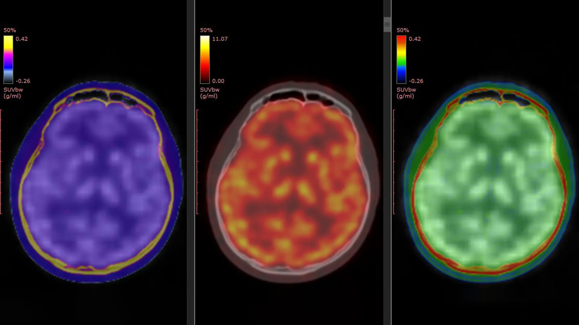
    The study, published in Molecular Psychiatry, used a PET tracer called [11C]K-2 designed to visualize AMPA receptors, proteins that help regulate communication between brain cells. Scientists have long suspected that AMPA receptor activity is central to ketamine’s antidepressant effects, but direct confirmation in living people has been limited.

    The researchers combined data from three clinical trials, comparing 34 patients with treatment-resistant depression against 49 healthy participants. Patients received intravenous ketamine or placebo over a two-week period, with PET scans taken before treatment and after the final infusion.

    Receptor shifts tied to symptom relief

    Before treatment, the PET data suggested patients with treatment-resistant depression had region-specific differences in AMPA receptor availability compared with healthy controls. After ketamine, the brain changes were not uniform, instead appearing as shifts in particular areas involved in mood and reward processing.

    Crucially, the degree and location of AMPA receptor changes tracked with how much a patient’s depressive symptoms improved. The authors highlighted especially notable shifts in regions linked in prior research to depression circuitry, arguing the images provide a direct bridge between earlier animal findings and human clinical response.

    Why it could matter clinically

    If replicated, AMPA receptor PET imaging could become a candidate biomarker to help predict who is most likely to benefit from ketamine, and to guide dosing or treatment strategies. That could be valuable because ketamine response can vary, and clinicians are seeking ways to personalize care while balancing benefit, side effects, and monitoring needs.

    The researchers caution that PET imaging is complex and not widely available, and larger studies would be needed before it could influence routine practice. Still, mapping ketamine’s effects at the receptor level may also support development of new rapid-acting antidepressants that target similar pathways with fewer practical barriers.

  • Tinnitus severity may be measurable at last: Study links pupil dilation and facial micro-movements to distress levels

    Researchers at Mass General Brigham say they have identified potential objective biomarkers for tinnitus severity by tracking pupil dilation and subtle, involuntary facial movements while people listen to everyday sounds. The work, published in Science Translational Medicine, aims to address a long-standing problem in tinnitus research: severity is typically judged by questionnaires rather than physiological measures.

    Tinnitus is commonly described as persistent phantom sound, such as ringing, buzzing, or clicking, and it is widespread in the general population. For many people it is a manageable nuisance, but a smaller group experiences debilitating distress that can disrupt sleep, concentration, and mental health.

    Signals tied to threat response

    The team focused on the sympathetic nervous system, which governs the body’s fight, flight, or freeze response, to look for outward indicators of distress. They examined whether tinnitus-related distress might be reflected in arousal signals that are visible in the eyes and face.

    To test the idea, researchers recruited 97 participants with normal hearing, including 47 with varying levels of tinnitus and sound sensitivity, and 50 control volunteers. Participants listened to pleasant, neutral, and unpleasant sounds while being recorded on video and monitored for pupil changes.

    AI helps detect tiny facial changes

    Using AI-powered video analysis, the study detected rapid micro-movements in areas such as the cheeks, eyebrows, and nostrils, and found they were associated with self-reported tinnitus distress. When these facial signals were combined with pupil dilation data, the model’s ability to predict severity improved.

    People with severe tinnitus showed unusually large pupil dilation across all sound types, while their facial responses were more muted. By contrast, controls and participants with less bothersome tinnitus tended to show stronger pupil and facial reactions mainly to the most unpleasant sounds.

    Why this could matter for trials

    The researchers argue that objective readouts could make placebo-controlled studies easier to design and interpret, helping the field evaluate treatments more rigorously. They also suggest the approach could potentially be adapted to more accessible, clinic-friendly tools if validated further.

    The team noted key limitations, including the need to exclude many people who often have complex tinnitus, such as those with hearing loss, older age, or significant mental health comorbidities. Future studies are expected to test whether the biomarkers hold up in broader, higher-risk populations and in real-world clinical settings.

    Researchers involved in the work say they are now exploring how these biomarkers could support therapy development, including approaches that pair neural stimulation with software-based treatment environments. The broader goal is to measure not just the sound people perceive, but the distress response that makes tinnitus disabling for some patients.