Blog

  • Reelin protein study hints at new way to strengthen leaky gut and reduce depression symptoms

    Reelin protein study hints at new way to strengthen leaky gut and reduce depression symptoms

    Researchers at the University of Victoria in Canada say a naturally occurring protein called Reelin may help explain how chronic stress can affect both the gut and the brain. Their findings, published in the journal Chronic Stress, point to Reelin as a potential target for future treatments aimed at gut barrier damage and depression-related symptoms.

    The study focuses on the gut barrier, the lining that controls what passes from the intestines into the bloodstream. Under prolonged stress, that barrier can become more permeable, a process often described as leaky gut, which may allow bacterial components to enter circulation and trigger immune inflammation.

    How stress may lower Reelin

    In preclinical experiments, the team observed that chronic stress was associated with reduced Reelin levels in intestinal tissue. When researchers administered a single small dose of Reelin, levels in the intestine returned to what the scientists described as normal ranges in their model.

    The authors argue the result matters because gut inflammation is increasingly studied as a contributor to mood disorders through the gut-brain axis. In this framework, immune signals and inflammatory processes can influence brain function, potentially worsening depression in vulnerable people.

    Why the gut-brain link matters

    Previous research has reported lower Reelin levels in brain tissue among people diagnosed with major depressive disorder, and similar patterns have been seen in stressed animal models. In earlier preclinical work cited by the team, Reelin administration was linked with antidepressant-like effects, though this does not yet demonstrate clinical benefit in humans.

    The researchers also highlight that the intestinal lining typically renews rapidly, roughly every few days, to maintain an effective barrier. They suggest Reelin may support this renewal process, which could help prevent barrier breakdown and reduce inflammation-driven effects that can intersect with depression.

    What comes next for Reelin research?

    Scientists caution that translating these findings into a therapy would require extensive follow-up, including safety studies, dosing work, and eventually human clinical trials. For now, the study adds to growing evidence that treating depression may, in some cases, involve addressing biological processes outside the brain, including gut integrity and immune activation.

    The research was supported by funding from Canada’s federal granting agencies, including the Canadian Institutes of Health Research and the Natural Sciences and Engineering Research Council of Canada. The authors say further studies will be needed to clarify how Reelin works across tissues and whether it can be developed into a practical medical intervention.

  • Cornell study suggests scent can predict platonic chemistry in minutes, and conversations may reshape what we smell

    New research from Cornell University suggests that scent plays a measurable role in how quickly strangers decide whether they could become friends. The study found that, within minutes, people’s reactions to another person’s everyday smell aligned with their impressions after a brief face-to-face chat.

    The work, published in Scientific Reports, examined how odor-based preferences interact with early social encounters. Researchers focused on friendship formation rather than romantic attraction, an area that has historically dominated social olfactory research.

    How the speed-friending test worked

    The study recruited heterosexual women who took part in four-minute speed-friending conversations. Participants also evaluated the everyday scent of other participants using T-shirts that had absorbed their normal, day-to-day odor.

    Researchers reported that individual, highly personal scent preferences predicted how much participants liked their interaction partners after the short conversations. Those patterns were not driven by a single universally appealing or unpleasant smell, but by idiosyncratic taste that varied by person.

    Everyday odor, not lab-made purity

    Instead of isolating a so-called natural body odor, the research leaned into what the authors describe as a signature scent shaped by daily life. That includes choices such as hygiene products, household environment, pets, and other common exposures that can influence how someone smells in real settings.

    In that sense, the study frames scent as part of a broader social signal people carry into first meetings, even if they are not consciously aware of it. The findings suggest smell is registered alongside conversation cues, body language, and other first-impression factors.

    Can a chat change how someone smells?

    One of the most notable findings was that the interaction appeared to work both ways. Participants’ ratings from the live conversation predicted shifts in how they later judged that same person’s T-shirt scent.

    That pattern suggests social experience can reshape odor perception, linking a person’s smell to the quality of the encounter. The researchers argue this feedback loop may help explain why people sometimes warm to someone over time, including on a sensory level.

    While the study focuses on a specific group and a controlled experimental design, it adds to evidence that scent influences social bonding beyond dating. The authors say future work could test whether similar effects appear across broader populations and in more natural social settings.

  • Penn State wearable sticker pairs biosensors and AI to spot genuine emotions, even behind a calm face

    Researchers at Penn State say they have developed a stretchable, rechargeable sticker designed to detect genuine emotions by combining facial movement data with physiological signals such as skin temperature and heart rate.

    The team argues the approach could help clinicians understand what patients feel in real time, especially when facial expressions alone are misleading or emotions are intentionally concealed.

    In a study published in Nano Letters, the researchers describe a BandAid-sized patch that measures several body signals linked to emotional states, including temperature, humidity, heart rate and blood oxygen levels.

    The device is built from thin, flexible layers of metals such as platinum and gold, shaped to remain sensitive even when bent, pulled or twisted during natural facial movement.

    How the emotion-tracking patch works

    To reduce measurement errors, the sensors are arranged so they operate independently, with protective layers intended to prevent stretching or moisture from distorting readings from neighboring components.

    Alongside the biosignals, facial strain sensors capture subtle changes in expression, and the system fuses those inputs to separate acted emotions from those tied to physiological responses.

    AI training and early accuracy results

    The researchers trained an AI model using repeated facial-expression performances across six categories: happiness, surprise, fear, sadness, anger and disgust.

    In the reported tests, the model classified performed facial expressions with 96.28% accuracy, based on data collected while participants repeatedly displayed each expression.

    To probe real emotions, participants watched video clips intended to elicit feelings while the patch tracked physiological changes associated with emotional arousal.

    The system identified emotions with 88.83% accuracy in those tests, with sensor readings aligning with known links between emotions and changes in metrics such as skin temperature and heart rate.

    Potential uses in telemedicine care

    The patch wirelessly transmits measurements to mobile devices and cloud systems, which the researchers say could support remote monitoring in telemedicine settings.

    The team also says the device is designed to avoid collecting personal information beyond sensor signals, aiming to reduce privacy risks while still enabling clinical interpretation.

    While the work remains at a research stage, the authors suggest the platform could eventually support broader health applications, including monitoring non-verbal patients and tracking conditions where behavioral signals are difficult to assess.

    The project was supported by funding from the U.S. National Institutes of Health and the U.S. National Science Foundation, according to the researchers.

  • MIT researchers map how focused ultrasound could test the brain circuits behind consciousness

    MIT researchers map how focused ultrasound could test the brain circuits behind consciousness

    Scientists at MIT are advancing plans to use transcranial focused ultrasound, a noninvasive technique that can modulate activity in deep brain regions, to study how conscious experience arises. The approach is laid out in a recent roadmap paper that argues the technology can move consciousness research beyond observation and toward direct tests of cause and effect.

    Consciousness remains a central unsolved problem in neuroscience because most tools either record brain signals or reach only surface areas without surgery. Focused ultrasound can concentrate acoustic energy through the skull onto small targets, potentially enabling precise stimulation of subcortical structures that are difficult to access with other noninvasive methods.

    A tool for cause and effect

    Many studies link conscious perception to patterns seen in EEG or brain imaging, but those signals often show correlation rather than causation. By changing neural activity in a controlled way and tracking what a person reports experiencing, researchers hope to identify which circuits are necessary for awareness and which are downstream side effects.

    MIT researchers Daniel Freeman and Matthias Michel, along with collaborators at the University of Florida and Harvard-affiliated Brigham and Women’s Hospital, argue that focused ultrasound could help narrow the search for the neural substrate of conscious perception. Their paper, published in Neuroscience and Biobehavioral Reviews, describes experimental designs intended for healthy volunteers.

    Testing rival theories of consciousness

    The roadmap highlights how the method could evaluate competing ideas about where consciousness is generated in the brain. Some accounts emphasize higher-level cognitive processes and the prefrontal cortex, while others suggest that specific perceptual regions, posterior networks, or deeper structures may be sufficient to generate conscious experience.

    Because the technology can target areas millimeters across, researchers say it may be possible to compare the effects of stimulating different nodes in these proposed networks. The goal is not only to see brain activity change, but to determine whether those changes reliably alter perception, awareness, or subjective reports.

    From vision to pain and beyond

    Early experiments are expected to start with the visual system, where researchers can tightly control stimuli and measure perception. Similar logic could be applied to pain, a domain where reflexive responses can occur before a person consciously feels discomfort, raising questions about which brain circuits produce the experience itself.

    While focused ultrasound has drawn growing interest for potential therapeutic uses, the authors frame it as a basic-science instrument for probing fundamental mechanisms. They also caution that, as with any emerging method, careful safety standards, calibration, and replication will determine how widely it can be adopted in mainstream neuroscience.

    At MIT, the work is part of a broader push to build a cross-disciplinary community around consciousness research, including regular discussions among neuroscientists and philosophers. For proponents, the appeal is straightforward: a noninvasive way to reach deeper brain targets could provide the most direct experimental leverage the field has had in decades.

  • Primary progressive aphasia study tests brain stimulation to boost speech therapy and what it could mean for patients

    Primary progressive aphasia, or PPA, is a neurodegenerative condition that gradually erodes a person’s ability to communicate, often affecting speech, writing, and word retrieval. There is currently no drug proven to halt or reverse the underlying progression, so care typically focuses on supportive speech-language therapy.

    Researchers at the University of Arizona are reporting encouraging results from an approach that pairs standard speech therapy with transcranial direct current stimulation, a noninvasive method that delivers a low electrical current through electrodes placed on the scalp. The goal is to amplify therapy gains by targeting brain networks involved in language.

    How the trial was designed

    The study, published in the Journal of Speech, Language, and Hearing Research, focused on logopenic PPA, a subtype often marked by difficulty finding words and repeating phrases. The team used neuroimaging to help identify stimulation targets while avoiding brain areas that were already significantly atrophied.

    Twelve participants with written language deficits completed two treatment phases in a randomized order, separated by a two-month break. In one phase, they received speech therapy plus active stimulation, and in the other, the same therapy paired with placebo stimulation.

    What improved after stimulation

    Participants improved after both phases, but the gains were stronger and lasted longer when active stimulation was added, the researchers reported. They observed clearer writing outcomes, including fewer spelling errors and better-formed, more meaningful sentences.

    Senior author Aneta Kielar said the team’s rationale reflects how language relies not only on meaning but also on retrieving a word’s sound structure during speaking and writing. Lead researcher Katlyn Nickels noted that PPA has only been widely characterized for several decades, leaving key aspects of treatment and recovery underexplored.

    Why neuroplasticity matters in PPA

    The researchers propose that stimulation may help promote neuroplasticity, the brain’s ability to reorganize and strengthen connections that support learning. In this view, brain stimulation does not replace therapy but may boost the effects of training by making language networks more responsive.

    They also emphasized that transcranial direct current stimulation is generally described in the field as relatively inexpensive and straightforward to administer under appropriate clinical protocols. Next, the group plans to study genetic, cognitive, and neural markers that could help predict which patients benefit most and how durable improvements can be in real-world care.

    The work was supported by multiple state and university health research programs, according to the authors. While larger studies are needed to confirm results and define best practices, the findings add to growing interest in combining rehabilitation with targeted neuromodulation for neurodegenerative language disorders.

  • Parkinson’s trial explores iPSC brain cell implants to restore dopamine and movement

    Parkinson’s trial explores iPSC brain cell implants to restore dopamine and movement

    Doctors at Keck Medicine of USC are taking part in an early-stage clinical trial testing whether implanted stem cell-derived brain cells can help people with Parkinson’s disease regain motor function. The approach aims to replace lost dopamine-producing cells and potentially reduce symptoms driven by dopamine decline.

    Parkinson’s is a progressive neurological condition that affects movement and can also influence mood and cognition. In the United States, more than 1 000 000 people live with the disease, and roughly 90 000 new cases are diagnosed each year, according to recent estimates.

    Aiming to restore dopamine production

    The trial focuses on replenishing dopamine, a key chemical messenger needed for smooth, coordinated movement. As dopamine-producing neurons deteriorate, people may develop tremor, muscle rigidity, slowed movement, and walking and balance difficulties.

    Standard therapies such as levodopa and deep brain stimulation can improve symptoms for many patients, but they do not replace the underlying lost cells. Researchers hope cell replacement could complement existing care by rebuilding the brain’s dopamine-making capacity.

    How the stem cell procedure works

    The study uses induced pluripotent stem cells, or iPSCs, which are adult cells reprogrammed into a flexible state and then guided to become dopamine-producing neurons. Because iPSCs are not embryonic stem cells, the technology is often presented as a less ethically contentious route for creating specialized cells.

    Neurosurgeons implant the cells into the basal ganglia using imaging guidance, aiming for precise placement in a region central to movement control. Participants are monitored closely for 12 to 15 months for safety signals and changes in Parkinson’s symptoms.

    Safety focus and limited enrollment

    Investigators are watching for risks that can follow brain surgery or cell therapies, including infection and abnormal involuntary movements known as dyskinesia. Longer follow-up is expected to continue for up to five years to better understand durability and longer-term safety.

    Keck Medicine is one of three U.S. sites participating in the Phase 1 REPLACE clinical trial, which plans to enroll 12 people with moderate to moderate-severe Parkinson’s disease. The experimental therapy, called RNDP-001, is being developed by Kenai Therapeutics and has received FDA fast-track designation to support an accelerated review pathway if results warrant it.

  • Study finds listeners use hand gestures to anticipate the next word in conversation

    In face-to-face conversation, people do more than listen to speech: they also read the hands. New research suggests that listeners use meaningful hand gestures to predict which words a speaker is likely to say next, speeding up comprehension.

    The work, led by scientists at the Max Planck Institute for Psycholinguistics and Radboud University in Nijmegen, tested whether gestures provide advance cues about upcoming speech. The team combined behavioural measures with electroencephalography, or EEG, to track how the brain responds.

    Avatars reveal predictive listening

    To tightly control timing and movement, the researchers used realistic virtual avatars that asked everyday questions. In one experiment, the avatar paused just before a key word, such as type, while making either a relevant typing gesture, a meaningless movement, or no movement at all.

    Participants were asked to guess how the sentence would end before hearing the missing word. They predicted the target word more often when they saw the matching gesture, indicating that hand movements can guide expectations about what comes next.

    EEG signals anticipation and easier processing

    A second experiment examined brain activity while a different group simply listened to the full questions. During the silent pause before the target word, EEG patterns associated with anticipation differed depending on whether a meaningful gesture was present.

    After the target word appeared, the brain showed a reduced N400 response when the gesture matched the word, a signal commonly linked to easier semantic processing. Together, the results suggest gestures help listeners prepare for upcoming meaning rather than merely adding emphasis after the fact.

    Implications for robots and assistants

    The findings also point to practical design choices for artificial agents, including robots and virtual assistants that use embodied avatars. If gestures can help humans predict speech in natural conversation, adding well-timed, meaningful hand movements could make synthetic communicators easier to understand.

    Researchers say the broader takeaway is that language comprehension is inherently multimodal. Listeners do not wait passively for words to arrive, but actively integrate visual cues such as gestures to anticipate what a speaker is about to say.

  • Cambridge study links menopause to grey matter decline, raising new questions about HRT and brain health

    Cambridge study links menopause to grey matter decline, raising new questions about HRT and brain health

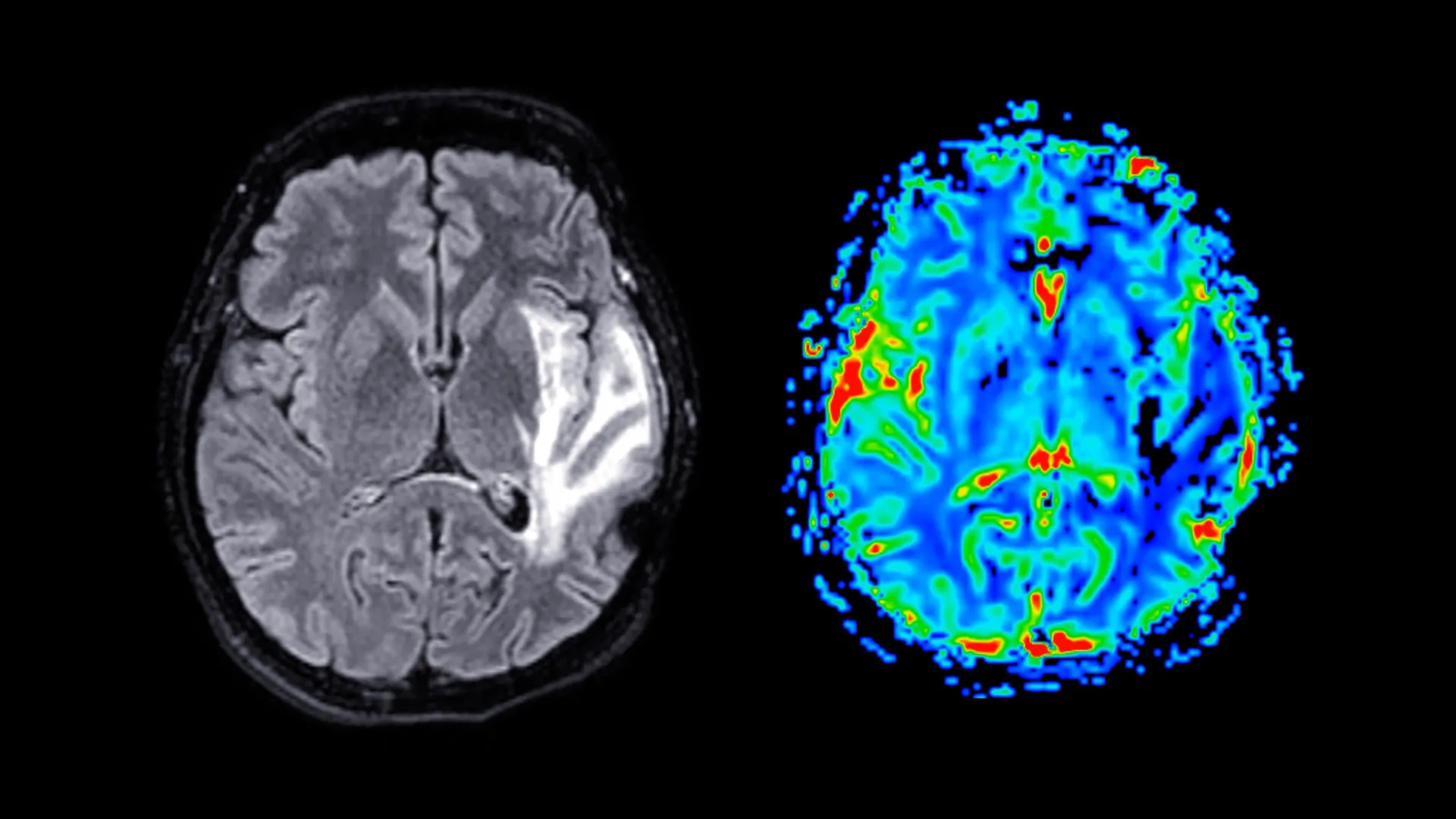
    Menopause may be associated with measurable changes in brain structure, alongside higher rates of anxiety, depression and sleep disruption, according to new research led by the University of Cambridge using UK Biobank data.

    In a large sample of women, researchers reported lower grey matter volume in several brain regions after menopause, patterns that were broadly similar whether or not participants had used hormone replacement therapy, commonly known as HRT.

    What the researchers analyzed

    The team examined questionnaire, health and cognitive testing data from nearly 125 000 women in the UK Biobank, a long-running project that links health records with detailed participant assessments.

    They also reviewed brain MRI scans from around 11 000 women, allowing comparisons between those who were pre-menopause, post-menopause without HRT use, and post-menopause with HRT use.

    Mental health and sleep symptoms

    Across the dataset, women who had gone through menopause were more likely to report seeking medical help for anxiety, nervousness or depression, and they were more likely to report persistent sleep problems such as insomnia and fatigue.

    Women who used HRT showed higher levels of anxiety and depression than non-users, but the analysis suggested these differences often existed before menopause, indicating HRT may have been prescribed to people already experiencing symptoms.

    Brain regions tied to memory and emotion

    Imaging results showed reduced grey matter volume after menopause in areas involved in memory and emotional regulation, including the hippocampus, entorhinal cortex and anterior cingulate cortex.

    Because some of these regions are also affected early in Alzheimer’s disease, the findings add to ongoing research into why women are diagnosed with dementia more often than men, though the study does not prove menopause causes dementia.

    On cognitive testing, memory scores were broadly similar across groups, but reaction time tended to be slower after menopause, with evidence that HRT use was associated with a smaller decline in reaction speed.

    The authors emphasized that menopause can be a major health transition and argued for greater attention to mental health support, sleep and lifestyle measures such as exercise and diet, alongside individualized medical advice about HRT.

    The study was published in Psychological Medicine, and the researchers noted that further work is needed to clarify how hormone changes, symptom severity, HRT timing and other health factors interact with brain ageing.

  • Study finds earlier bedtimes and longer sleep may sharpen teens cognitive performance, even with small differences

    Adolescents who sleep a little longer and tend to fall asleep earlier show stronger brain function and do better on cognitive tests than peers with later, shorter sleep, according to researchers in the UK and China.

    The findings draw on objective sleep tracking and brain imaging, offering fresh evidence that modest changes in sleep habits may be linked to measurable differences in how the teenage brain works.

    Researchers analyzed data from the Adolescent Brain Cognitive Development study in the United States, using Fitbit sleep measures from more than 3 200 participants aged 11 to 12 and comparing them with brain scans and cognitive testing.

    They then checked whether similar patterns appeared in two additional groups aged 13 to 14, totaling about 1 190 participants, to see if the results held up beyond a single age snapshot.

    The team identified three broad sleep profiles, with average sleep times ranging from about 7 hours 10 minutes to roughly 7 hours 25 minutes, a gap of just over 15 minutes between the shortest and longest sleepers.

    Despite the small difference, adolescents in the longest-sleep group performed best on tests that assess skills such as vocabulary, reading, problem solving, and focus.

    Brain measures differed across groups

    Brain imaging also showed differences that tracked with sleep patterns, with the longest-sleep group showing the largest overall brain volume and stronger brain function measures, while the shortest-sleep group showed the smallest volume and weakest measures.

    The study did not find significant differences in school achievement between groups, suggesting standardized academic outcomes may not capture the subtler cognitive effects observed in testing.

    Heart-rate data during sleep pointed in the same direction: the longest-sleep group had the lowest heart rates across sleep states, while the shortest-sleep group had the highest.

    Lower sleeping heart rates are generally associated with better cardiovascular health and can align with more stable, higher-quality sleep, while higher rates can accompany restless sleep and frequent awakenings.

    Most teens still fell short

    Even the best sleepers in the study were not reaching the amount of sleep typically recommended for adolescents, highlighting how widespread sleep shortfalls can be in early teen years.

    The American Academy of Sleep Medicine advises that teenagers aged 13 to 18 should regularly sleep 8 to 10 hours per night for optimal health, while many fall below that range.

    Because the dataset follows participants over time, researchers reported that differences in sleep patterns and related brain and cognitive measures appeared to persist across multiple years around the main assessment window.

    The authors cautioned that the study cannot prove that better sleep directly causes better brain function, but they noted prior research supporting sleep’s role in memory consolidation and learning.

    What could be driving later bedtimes?

    The researchers said the next step is to better understand why some adolescents consistently go to bed later and sleep less, including potential influences such as evening screen use and individual body-clock differences.

    They argue that identifying the drivers of sleep loss could help shape practical interventions, since the results suggest even small improvements in sleep timing and duration may matter.

  • Why astringent flavanols in cocoa and berries may help trigger brain activity

    Why astringent flavanols in cocoa and berries may help trigger brain activity

    A dry, puckering sensation from cocoa, some berries and red wine is more than a taste quirk, according to emerging research on flavanols. Scientists are increasingly investigating whether astringency itself can act as a rapid signal to the brain, potentially influencing attention and memory.

    Flavanols are a type of polyphenol long associated in population studies and clinical research with cardiovascular benefits, including improved blood vessel function. They have also been linked to cognitive outcomes, but one persistent challenge is that only a small fraction of consumed flavanols is absorbed into the bloodstream.

    A new focus on taste pathways

    In a recent study in Current Research in Food Science, researchers from Shibaura Institute of Technology in Japan proposed that the sensory experience of astringency may help explain flavanols’ outsized effects. The team hypothesized that stimulation in the mouth could transmit signals through sensory nerves to the central nervous system.

    Working with mice, the researchers administered oral doses of flavanols and compared results with a control group given water. The flavanol groups showed higher activity and exploratory behavior and performed better on learning and memory tasks in the experiments.

    Neurochemistry tied to alertness and stress

    Brain measurements suggested changes in neurotransmitter systems associated with attention and arousal, including dopamine-related activity and the locus coeruleus norepinephrine network. The study also reported shifts in markers linked to sympathetic nervous system activity, which plays a key role in alertness and the body’s stress response.

    The researchers interpreted these patterns as evidence that flavanol-driven astringency may function like a mild physiological challenge, with downstream effects that resemble some aspects of exercise-induced activation. They argue this could help reconcile low bioavailability with observed impacts on brain-related outcomes.

    What it means for everyday diets

    The findings do not mean that any bitter or drying food will reliably boost cognition, and the work is primarily an animal study rather than a clinical trial in humans. Still, it adds momentum to the broader scientific push to understand how sensory cues from food can influence the brain quickly, alongside longer-term effects from digestion and circulation.

    Researchers say the idea could inform future work in sensory nutrition, including how foods might be formulated to balance palatability with measurable physiological responses. For consumers, the most evidence-backed approach remains obtaining flavanol-rich foods as part of an overall healthy diet, rather than treating astringency as a standalone brain hack.