Blog

  • MIT’s ComMAND gene circuit could make gene therapy dosing safer and more predictable

    Gene therapy has long promised one-time treatments for disorders caused by a missing or faulty gene, but controlling how strongly a delivered gene turns on in cells remains a major hurdle. Too little expression can leave a therapy ineffective, while too much can raise toxicity and other safety risks.

    Engineers at MIT report a compact gene-control design that aims to keep expression within a targeted range, even when cells receive different numbers of gene copies. The work, published in Cell Systems, centers on a circuit the team calls ComMAND, short for Compact microRNA-mediated attenuator of noise and dosage.

    A built-in brake for expression

    Many gene therapies rely on viral vectors such as adeno-associated virus or lentivirus to deliver therapeutic DNA. But uptake varies widely from cell to cell, which can create large swings in how much protein a new gene produces.

    ComMAND uses a control strategy known as an incoherent feedforward loop, pairing gene activation with a simultaneous suppressor signal. In this design, the therapeutic gene also produces a microRNA that dampens its own translation, acting as an internal counterweight.

    Compact design fits common vectors

    The researchers engineered the microRNA sequence inside an intron within the therapeutic gene, so both the gene’s messenger RNA and the suppressing microRNA are produced together. That single-transcript setup is intended to smooth out variability when delivery levels differ across cells.

    Because the circuit can be controlled with one promoter, the team says expression can be tuned by selecting promoters of different strengths. The compact architecture is also designed to fit within a single delivery vehicle, which could simplify manufacturing and development.

    Early results across multiple cell types

    In human cells, the team demonstrated ComMAND with genes linked to Friedreich’s ataxia and Fragile X syndrome, aiming to keep expression closer to desired levels. They reported gene output around eight times typical healthy levels in their tests, compared with more than 50 times without the circuit.

    The approach was also evaluated in rat neurons, mouse fibroblasts, and human T-cells using a fluorescent reporter to measure expression. The researchers say the next step is to test whether this tighter control can restore function and improve disease signs in cultured systems and animal models.

    The authors note that many candidate conditions are rare, making it difficult to run large studies and optimize dosing. They argue that more predictable, tunable gene circuits could lower development barriers for therapies targeting small patient populations.

  • AI study finds stroke may make the opposite brain hemisphere look younger, offering new clues on recovery

    AI study finds stroke may make the opposite brain hemisphere look younger, offering new clues on recovery

    A new study in The Lancet Digital Health suggests the brain can respond to stroke in a surprising way. Researchers at the USC Mark and Mary Stevens Neuroimaging and Informatics Institute (Stevens INI) found that people with severe physical impairments after a stroke may show signs of a “younger” brain structure in areas that were not damaged. This appears to reflect how the brain adapts and reorganizes itself after injury.

    The research was conducted as part of the Enhancing NeuroImaging Genetics through Meta-Analysis (ENIGMA) Stroke Recovery Working Group. Scientists analyzed brain scans from more than 500 stroke survivors collected across 34 research centers in eight countries. By applying deep learning models trained on tens of thousands of MRI scans, the team estimated the “brain age” of different regions in each hemisphere and examined how stroke affects both structure and recovery.

    “We found that larger strokes accelerate aging in the damaged hemisphere but paradoxically make the opposite side of the brain appear younger,” said Hosung Kim, PhD, associate professor of research neurology at the Keck School of Medicine of USC and co-senior author of the study. “This pattern suggests the brain may be reorganizing itself, essentially rejuvenating undamaged networks to compensate for lost function.”

    AI Reveals Brain Rewiring After Stroke

    To carry out the analysis, researchers used a type of artificial intelligence called a graph convolutional network. This system estimated the biological age of 18 brain regions based on MRI data. They then compared this predicted age with each person’s actual age, a measure known as the brain-predicted age difference (brain-PAD), which serves as an indicator of brain health.

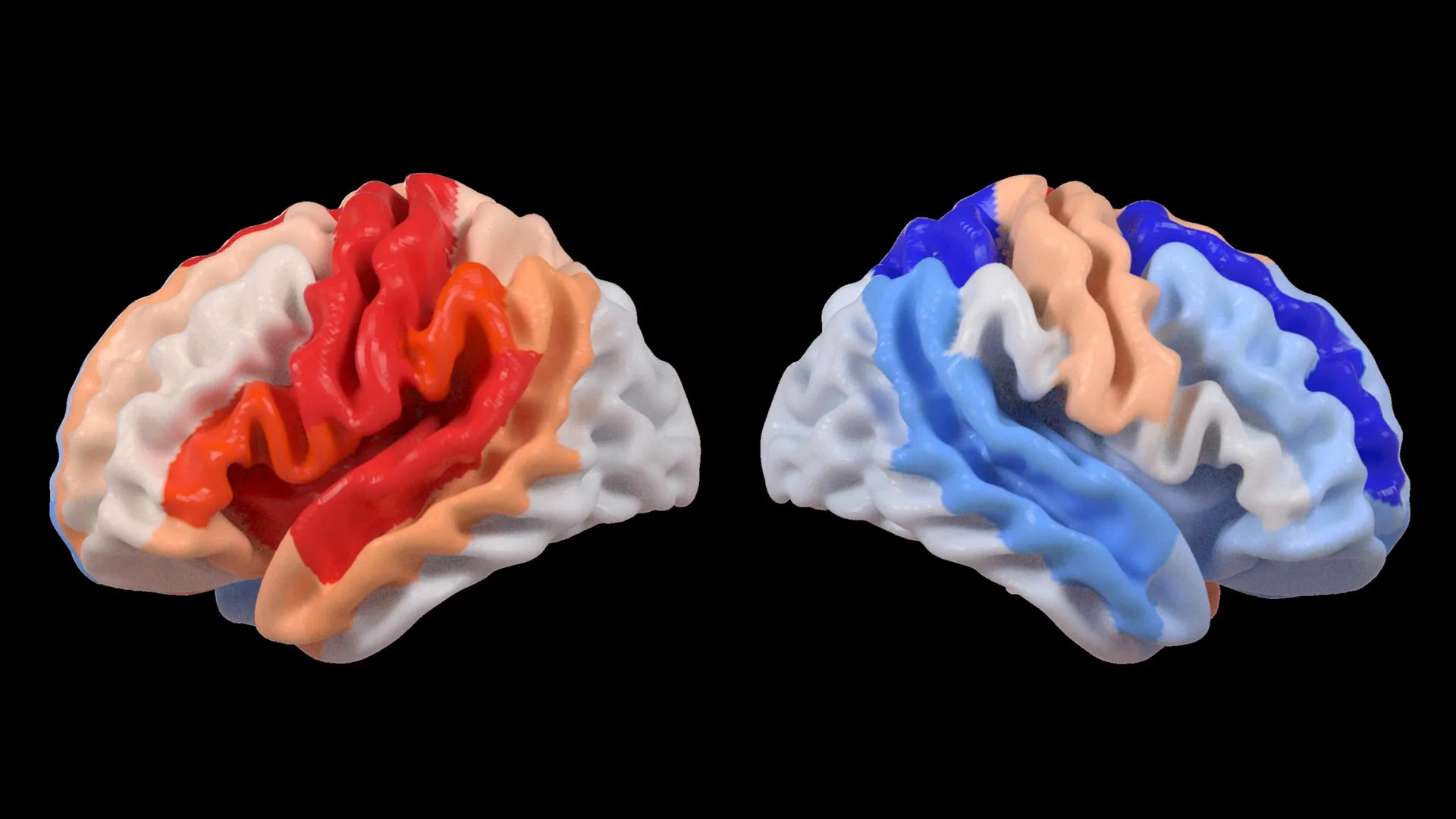
    When these brain age measurements were compared with motor function scores, a clear pattern emerged. Stroke survivors with severe movement impairments, even after more than 6 months of rehabilitation, showed younger-than-expected brain age in regions opposite the site of injury. This effect was especially strong in the frontoparietal network, which plays an important role in movement planning, attention, and coordination.

    “These findings suggest that when stroke damage leads to greater movement loss, undamaged regions on the opposite side of the brain may adapt to help compensate,” Kim explained. “We saw this in the contralesional frontoparietal network, which showed a more ‘youthful’ pattern and is known to support motor planning, attention, and coordination. Rather than indicating full recovery of movement, this pattern may reflect the brain’s attempt to adjust when the damaged motor system can no longer function normally. This gives us a new way to see neuroplasticity that traditional imaging could not capture.”

    Large-Scale Data Reveals Hidden Patterns

    The study relied on ENIGMA, a global collaboration that combines data from more than 50 countries to better understand the brain across different conditions. By standardizing MRI data and clinical information from many research groups, the team created the largest stroke neuroimaging dataset of its kind.

    “By pooling data from hundreds of stroke survivors worldwide and applying cutting-edge AI, we can detect subtle patterns of brain reorganization that would be invisible in smaller studies. These findings of regionally differential brain aging in chronic stroke could eventually guide personalized rehabilitation strategies,” said Arthur W. Toga, PhD, director of the Stevens INI and Provost Professor at USC.

    Toward Personalized Stroke Recovery

    The researchers plan to continue this work by following patients over time, from the early stages after a stroke through long-term recovery. Tracking how brain aging patterns and structural changes evolve could help doctors tailor treatments to each person’s unique recovery process, with the goal of improving outcomes and quality of life.

    Learn more about associations between contralesional neuroplasticity and motor impairment by viewing this video made by the Stevens INI.

    The study, “Deep learning prediction of MRI-based regional brain age reveals contralesional neuroplasticity associated with severe motor impairment in chronic stroke: A worldwide ENIGMA study,” was funded by the National Institutes of Health (NIH) grant R01 NS115845 and supported by international collaborators from institutions including the University of British Columbia, Monash University, Emory University, and the University of Oslo.

  • Hong Kong PolyU researchers unveil AI tool to score large language model personality, with implications for business compliance and education

    Large language models have become a default interface for many AI products, but researchers still struggle to describe their behaviour in a consistent, measurable way. A team at The Hong Kong Polytechnic University says it has built a system that aims to quantify an LLM’s personality based on linguistic output.

    The tool, called Language Model Linguistic Personality Assessment, or LMLPA, is designed to translate model responses into numerical scores tied to personality traits. The researchers describe it as a step toward making model behaviour easier to compare across systems and deployments.

    How the LMLPA system works

    LMLPA combines two components: an adapted version of the Big Five Inventory and an AI rater that grades the model’s answers. The process focuses on patterns such as wording, style and other language features found in generated text.

    By using a standardised questionnaire structure, the approach attempts to bring more consistency to assessments that often rely on subjective impressions. The researchers say the outcome is a data-driven profile that can be tracked and tested across different prompts and settings.

    Why personality metrics could matter

    Developers and organisations increasingly want AI assistants to behave predictably in sensitive contexts, including classrooms, customer support and internal decision workflows. The team argues that quantifying communication tendencies could help tailor a model’s tone and interaction style to a specific use case.

    The researchers also point to potential value in governance and oversight, where firms are under pressure to document how AI tools behave and how risks are managed. They suggest that structured behavioural metrics could complement existing evaluation methods focused on accuracy and safety.

    From research to compliance applications

    PolyU said the work has also informed a business compliance platform that uses natural language processing to analyse large volumes of reports and other text. In that context, automation is intended to speed up data collection, analysis and insight generation for reporting tasks.

    The study, led by Prof. Lik-Hang Lee of PolyU’s Department of Industrial and Systems Engineering, was published in the journal Computational Linguistics. The researchers position LMLPA as part of a broader effort to align AI systems with human values and practical operational needs.

  • PET brain scans map ketamine’s rapid antidepressant effect, pointing to a potential biomarker for treatment-resistant depression

    PET brain scans map ketamine’s rapid antidepressant effect, pointing to a potential biomarker for treatment-resistant depression

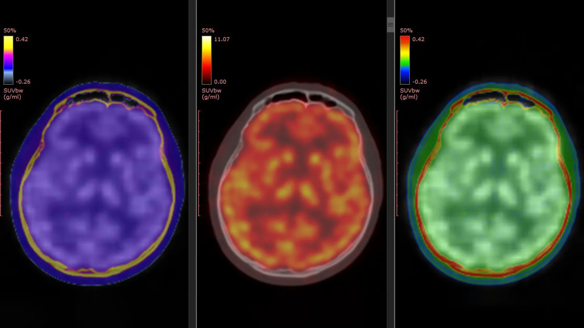
    Researchers in Japan have reported some of the clearest human evidence yet of how ketamine can relieve symptoms of treatment-resistant depression, using PET brain imaging to track changes in key glutamate receptors. The work adds molecular detail to a treatment already known for acting faster than standard antidepressants.

    Major depressive disorder is a leading cause of disability worldwide, and a substantial share of patients do not improve after trying multiple first-line therapies. For those with treatment-resistant depression, ketamine and the related medicine esketamine have drawn attention because some patients feel relief within hours or days rather than weeks.

    What the PET scans measured

    The study, published in Molecular Psychiatry, used a PET tracer called [11C]K-2 designed to visualize AMPA receptors, proteins that help regulate communication between brain cells. Scientists have long suspected that AMPA receptor activity is central to ketamine’s antidepressant effects, but direct confirmation in living people has been limited.

    The researchers combined data from three clinical trials, comparing 34 patients with treatment-resistant depression against 49 healthy participants. Patients received intravenous ketamine or placebo over a two-week period, with PET scans taken before treatment and after the final infusion.

    Receptor shifts tied to symptom relief

    Before treatment, the PET data suggested patients with treatment-resistant depression had region-specific differences in AMPA receptor availability compared with healthy controls. After ketamine, the brain changes were not uniform, instead appearing as shifts in particular areas involved in mood and reward processing.

    Crucially, the degree and location of AMPA receptor changes tracked with how much a patient’s depressive symptoms improved. The authors highlighted especially notable shifts in regions linked in prior research to depression circuitry, arguing the images provide a direct bridge between earlier animal findings and human clinical response.

    Why it could matter clinically

    If replicated, AMPA receptor PET imaging could become a candidate biomarker to help predict who is most likely to benefit from ketamine, and to guide dosing or treatment strategies. That could be valuable because ketamine response can vary, and clinicians are seeking ways to personalize care while balancing benefit, side effects, and monitoring needs.

    The researchers caution that PET imaging is complex and not widely available, and larger studies would be needed before it could influence routine practice. Still, mapping ketamine’s effects at the receptor level may also support development of new rapid-acting antidepressants that target similar pathways with fewer practical barriers.

  • Tinnitus severity may be measurable at last: Study links pupil dilation and facial micro-movements to distress levels

    Researchers at Mass General Brigham say they have identified potential objective biomarkers for tinnitus severity by tracking pupil dilation and subtle, involuntary facial movements while people listen to everyday sounds. The work, published in Science Translational Medicine, aims to address a long-standing problem in tinnitus research: severity is typically judged by questionnaires rather than physiological measures.

    Tinnitus is commonly described as persistent phantom sound, such as ringing, buzzing, or clicking, and it is widespread in the general population. For many people it is a manageable nuisance, but a smaller group experiences debilitating distress that can disrupt sleep, concentration, and mental health.

    Signals tied to threat response

    The team focused on the sympathetic nervous system, which governs the body’s fight, flight, or freeze response, to look for outward indicators of distress. They examined whether tinnitus-related distress might be reflected in arousal signals that are visible in the eyes and face.

    To test the idea, researchers recruited 97 participants with normal hearing, including 47 with varying levels of tinnitus and sound sensitivity, and 50 control volunteers. Participants listened to pleasant, neutral, and unpleasant sounds while being recorded on video and monitored for pupil changes.

    AI helps detect tiny facial changes

    Using AI-powered video analysis, the study detected rapid micro-movements in areas such as the cheeks, eyebrows, and nostrils, and found they were associated with self-reported tinnitus distress. When these facial signals were combined with pupil dilation data, the model’s ability to predict severity improved.

    People with severe tinnitus showed unusually large pupil dilation across all sound types, while their facial responses were more muted. By contrast, controls and participants with less bothersome tinnitus tended to show stronger pupil and facial reactions mainly to the most unpleasant sounds.

    Why this could matter for trials

    The researchers argue that objective readouts could make placebo-controlled studies easier to design and interpret, helping the field evaluate treatments more rigorously. They also suggest the approach could potentially be adapted to more accessible, clinic-friendly tools if validated further.

    The team noted key limitations, including the need to exclude many people who often have complex tinnitus, such as those with hearing loss, older age, or significant mental health comorbidities. Future studies are expected to test whether the biomarkers hold up in broader, higher-risk populations and in real-world clinical settings.

    Researchers involved in the work say they are now exploring how these biomarkers could support therapy development, including approaches that pair neural stimulation with software-based treatment environments. The broader goal is to measure not just the sound people perceive, but the distress response that makes tinnitus disabling for some patients.

  • Study links early depression to brain cell energy changes, hinting at a future blood test

    Study links early depression to brain cell energy changes, hinting at a future blood test

    New research suggests major depressive disorder may be tied to early disruptions in how cells generate and manage energy, a finding that could eventually support earlier and more targeted treatment. Scientists say the results add biological detail to a condition still often diagnosed mainly through symptoms and clinical interviews.

    The study focused on adenosine triphosphate, or ATP, sometimes described as the body’s energy currency because it powers basic cellular work. Researchers examined ATP-related signals in both the brain and blood, aiming to see whether measurable energy patterns track with depression in young adults.

    What the scientists measured

    Teams at the University of Queensland and the University of Minnesota analyzed brain imaging and blood samples from 18 participants aged 18 to 25 diagnosed with major depressive disorder. Their results were compared with samples from people without depression to identify differences linked to the illness.

    According to the researchers, the approach is notable because it looked for matching patterns across the brain and the bloodstream, not just in one system. That raises the possibility that, with more evidence, peripheral markers in blood could one day help flag risk or subtypes of depression earlier.

    An unexpected pattern under stress

    The researchers reported that cells from participants with depression showed higher energy-molecule production while at rest, but had difficulty ramping up energy output when challenged. That stress-response limitation, they argue, could align with common symptoms such as fatigue, slowed thinking, and reduced motivation.

    Scientists involved in the work suggest the pattern may reflect mitochondria that are effectively overcompensating early on, then struggling when demand increases. They caution that the study is small, but say it offers a plausible cellular mechanism worth testing in larger groups.

    What this could mean next

    Major depressive disorder is common and can take years to match with an effective treatment, particularly when fatigue is prominent and persistent. The authors argue that identifying measurable biological signatures could support earlier intervention and more personalized care, rather than trial-and-error alone.

    The research was published in Translational Psychiatry, and the team says follow-up studies are needed to confirm the findings, test whether they predict outcomes, and determine whether they apply across ages and different forms of depression. If replicated, the work could also help frame depression as a whole-body condition with detectable biological changes.

  • Microplastics and the brain: Researchers map possible links to Alzheimer’s and Parkinson’s

    Microplastics and the brain: Researchers map possible links to Alzheimer’s and Parkinson’s

    Microplastics, the tiny plastic fragments found in food, water and household dust, are under growing scrutiny as researchers examine how they may affect the brain. A new scientific review pulls together evidence suggesting these particles could contribute to processes seen in Alzheimer’s and Parkinson’s disease.

    Dementia affects more than 57 million people globally, and experts expect the burden of neurodegenerative disease to rise as populations age. That backdrop is sharpening interest in whether environmental exposures such as microplastics may worsen inflammation or accelerate neurological decline.

    Five mechanisms under scientific review

    The review, published in Molecular and Cellular Biochemistry by an international team including the University of Technology Sydney and Auburn University, outlines several biological routes of potential harm. It focuses on immune activation, oxidative stress, disruption of the blood-brain barrier, mitochondrial dysfunction and direct neuronal injury.

    Researchers argue that if microplastics weaken the blood-brain barrier, the brain may become more vulnerable to inflammatory molecules and immune responses that can damage delicate tissue. In parallel, they describe how oxidative stress could rise if reactive oxygen species increase while antioxidant defenses are depleted.

    Energy disruption and protein buildup concerns

    Another concern highlighted in the paper is mitochondrial interference, which could reduce cellular energy production and strain neurons that rely heavily on steady ATP supply. Over time, energy shortfalls may impair brain function and make nerve cells more susceptible to damage.

    The authors also discuss disease-specific hypotheses, including whether microplastics might promote protein changes associated with Alzheimer’s, such as beta-amyloid and tau accumulation. For Parkinson’s, they note a possible role in α-synuclein aggregation and stress on dopamine-producing neurons.

    What the evidence can and cannot show

    While the review raises plausible pathways, the researchers stress that confirming a direct causal link in humans will require further studies, including exposure measurement and long-term clinical follow-up. Much of the current understanding comes from laboratory and animal research, along with emerging evidence that microplastics can accumulate in organs.

    Even with uncertainties, scientists say practical exposure reduction may be reasonable while research catches up, particularly in everyday food and household contexts. The authors point to reducing reliance on plastic food containers and packaging, limiting plastic-related dust and fibers, and supporting policies that curb plastic pollution at its source.

    Ongoing work at the involved institutions is expected to further test how ingested or inhaled microplastics interact with cells and barriers in the body. Public health experts say clearer answers will depend on standardizing how microplastics are measured and comparing real-world doses across populations.

  • Explainable AI tool CANYA decodes protein aggregation patterns, offering new clues for amyloid diseases and drug manufacturing

    An AI tool has made a step forward in translating the language proteins use to dictate whether they form sticky clumps similar to those linked to Alzheimer’s Disease and around fifty other types of human disease. In a departure from typical “black-box” AI models, the new tool, CANYA, was designed to be able to explain its decisions, revealing the specific chemical patterns that drive or prevent harmful protein folding.

    The discovery, published today in the journal Science Advances, was possible thanks to the largest-ever dataset on protein aggregation created to date. The study gives new insights about the molecular mechanisms underpinning sticky proteins, which are linked to diseases affecting half a billion people worldwide.

    Protein clumping, or amyloid aggregation, is a health hazard that disrupts normal cell function. When certain patches in proteins stick to each other, proteins grow into dense fibrous masses that have pathological consequences.

    While the study has some implications for accelerating research efforts for neurodegenerative diseases, it’s more immediate impact will be in biotechnology. Many drugs are proteins, and they are often hampered by unwanted clumping.

    “Protein aggregation is a major headache for pharmaceutical companies,” says Dr. Benedetta Bolognesi, co-corresponding author of the study and Group Leader at the Institute for Bioengineering of Catalonia (IBEC).

    “If a therapeutic protein starts aggregating, manufacturing batches can fail, costing time and money. CANYA can help guide efforts to engineer antibodies and enzymes that are less likely to stick together and reduce expensive setbacks in the process,” she adds.

    Protein clumps are formed using a poorly understood language. Proteins are made of twenty different types of amino acids. Instead of the usual A, C, G, T letters that make up the language of DNA, a protein’s language has twenty different letters, different combinations of which form “words” or “motifs.”

    Researchers have long sought to decipher which combinations of motifs cause clumping and which others enable proteins to fold without error. Artificial intelligence tools that treat amino acids like the alphabet of a mysterious language could help identify the precise words or motifs responsible, but the quality and volume of data about protein aggregation needed to feed models have been historically scant or restricted to very small protein fragments.

    The study addressed this challenge by carrying out large-scale experiments. The authors of the study created over 100,000 completely random protein fragments, each 20 amino acids long, from scratch. The ability for each synthetic fragment to clump was tested in living yeast cells. If a particular fragment triggered clump formation, the yeast cells would grow in a certain way that could be measured by the researchers to determine cause and effect.

    Around one in every five protein fragments (21,936/100,000) caused clumping, while the rest did not. While previous studies might have tracked a handful sequences, the new dataset captures a much bigger catalogue of the different protein variants which can cause amyloid aggregation.

    “We created truly random protein fragments including many versions not found in nature. Evolution has explored only a fraction of all possible protein sequences, while our approach helps us peer into a much bigger galaxy of possibilities, providing lots of data points to help understand more general laws of aggregation behaviour,” explains Dr. Mike Thompson, first author of the study and postdoctoral researcher at the Centre for Genomic Regulation (CRG).

    The vast amount of data generated from the experiments was used to train CANYA. The researchers decided to create it using the principles of “explainable AI,” making its decision-making processes transparent and understandable to humans. This meant sacrificing a little bit of its predictive power, which is usually higher in “black-box” AIs. Despite this, CANYA proved to be around 15% more accurate than existing models.

    Specifically, CANYA is a convolution-attention model, a hybrid tool borrowing from two distinct corners of AI. Convolution models, like those used in image recognition, scan photos for features like an ear or a nose to identify a face, except in this case CANYA skims through the protein chain to find meaningful features like motifs or “words.”

    Attention AI models are used by language translation tools to identify key phrases in a sentence before deciding on the best translation. The researchers incorporated this technique to help CANYA figure out which motifs matter most in the grand scheme of the entire protein.

    Together, these two approaches help CANYA see local motifs up close while also spotting their bigger-picture importance. The researchers could use this information to not just predict which motifs in the protein chain encourage clumping, block it, or something in between, but also understand why.

    For example, CANYA showed that small pockets of water-repelling amino acids are more likely to spark clumping, while some motifs have a bigger impact on clumping if they’re near the start of a protein sequence rather than at the end. The observations align with previous findings researchers have seen under the microscope in known amyloid fibrils.

    But CANYA also found new rules driving protein aggregation. For instance, certain building blocks of proteins, so-called charged amino acids, are normally thought to prevent clumping. But it turns out that in the context of other specific building blocks, they can actually promote clumping.

    In its current form, CANYA primarily explains protein aggregation in yes or no terms, i.e. it works as a so-called “classifier.” The researchers next want to refine the system so it can predict and compare aggregation speeds rather than just aggregation likelihood. This could help predict which protein variants form clumps quickly and which do so more slowly, a vital factor in neurodegenerative diseases where the timing of amyloid formation matters just as much as the fact that it happens at all.

    “There are 1024 quintillion ways of creating a protein fragment that is 20-amino acids long. So far, we’ve trained an AI with just 100,000 fragments. We want to improve it by making more and bigger fragments. This is just the first step but our work shows it is possible to decipher the language of protein aggregation. This is incredibly important for our understanding of human disease but also to guide synthetic biology efforts” concludes Dr. Bolognesi.

    “This project is a great example of how combining large-scale data generation with AI can accelerate research. It’s also a very cost-effective method to generate data,” says ICREA Research Professor Ben Lehner, co-corresponding author and Group Leader at the Centre for Genomic Regulation (CRG) and the Wellcome Sanger Institute.

    “Using DNA synthesis and sequencing we can perform hundreds of thousands of experiments in a single tube, generating the data we need to train AI models. This is an approach we are applying to many difficult problems in biology. The goal is to make biology predictable and programmable,” he adds.

    The study is a joint collaborative effort by ICREA Research Professor Ben Lehner’s lab at the Centre for Genomic Regulation (CRG) and Benedetta Bolognesi’s lab at the Institute for Bioengineering of Catalonia (IBEC). Researchers from Cold Spring Harbor Laboratory (CSHL) and Wellcome Sanger Institute also collaborated in the study. It was funded by “La Caixa” Research Foundation, the European Research Council and the Spanish Ministry of Science and Innovation.
  • Teen Diet and Depression Risk: A Major Review Points to Whole Foods Over Supplements

    Teen Diet and Depression Risk: A Major Review Points to Whole Foods Over Supplements

    A major review led by Swansea University researchers suggests teenagers’ overall diet quality may be linked to mental health, with healthier eating patterns more often associated with fewer depressive symptoms. The findings add to growing interest in nutrition as a modifiable factor that could support adolescent wellbeing.

    Published in the journal Nutrients, the paper assessed evidence from 19 earlier studies examining diet and mental health in adolescents. Across the studies, lower-quality diets were more frequently connected with higher psychological distress, though results varied by design and population.

    Whole-diet patterns stand out

    The review included six randomized controlled trials and 13 prospective cohort studies, allowing the authors to compare different types of evidence. While some studies hinted that specific supplements such as vitamin D might reduce depressive symptoms, the overall picture for individual nutrients was inconsistent.

    By contrast, broader dietary patterns showed clearer and more repeatable signals. The authors argue that focusing on overall balance and quality may be more useful than targeting single nutrients, particularly when translating research into school, family, and public health settings.

    Why adolescence is a key window

    Researchers highlighted adolescence as a critical period for brain development and emotional regulation, when depression symptoms can emerge and become entrenched. Because diet is part of daily life and can be shaped at scale, they see it as a practical area for prevention and early support.

    At the same time, the review notes that diet and mental health do not exist in isolation. Socioeconomic factors and sex differences may influence both what teens eat and how mental health outcomes present, complicating cause-and-effect interpretation.

    What the evidence still misses

    The authors flagged major gaps, including a heavy focus on depression compared with other outcomes such as anxiety, stress, self-esteem, externalizing behavior, and aggression. They also emphasized the need for more standardized methods and improved reporting so results can be compared across studies.

    To strengthen future conclusions, the team proposed a research roadmap that includes exposure-based designs, biological markers, and open science practices. In a statement, corresponding author Hayley Young said, “Overall, our findings suggest that public health and clinical strategies should prioritise whole-diet approaches over isolated supplementation when considering adolescent mental health.”

    The researchers cautioned that more high-quality studies are needed to determine which dietary patterns work best, and for whom. Even so, the review suggests that everyday food choices may play a bigger role in teen mental health than many families and clinicians have assumed.

  • University of Tokyo Study Finds Motor Exploration Builds a Stronger Sense of Agency in New Human-Computer Tasks

    Why do people feel they are in control of their own movements, especially when learning an unfamiliar task? New research from the University of Tokyo examines how the brain builds a sense of agency, the feeling that an action and its outcome are self-generated.

    Sense of agency is central to everyday activities, from walking and typing to manipulating objects in the environment. It is also increasingly relevant to modern human-computer interfaces, including virtual reality tools, assistive devices, and emerging brain-machine technologies.

    How the brain detects control

    Researchers often explain agency using the comparator model, where the brain predicts what should happen when a person moves and then compares that prediction with incoming sensory feedback. When prediction and feedback align, the feeling of control tends to strengthen.

    The Tokyo team noted a gap in this framework when applied to learning from scratch, when accurate predictions do not yet exist. In early learning, people often try actions first and only later infer the rules linking movement to outcomes.

    Testing agency during new motor learning

    In the study, participants used a specialized data glove to move a cursor on a screen through finger motions, learning the hand-to-screen mapping by trial and error. At different stages, the researchers assessed how strongly participants felt they controlled the cursor, including when the cursor was subtly shifted in space or delayed in time.

    During the earliest stage, participants relied heavily on timing, judging agency largely by whether the cursor moved in sync with their fingers. With practice, agency increasingly depended on whether the cursor followed the learned mapping, a pattern that was strongest among higher-performing participants.

    Why exploration mattered

    A second experiment reduced motor exploration by having participants imitate presented gestures aimed at reaching target positions. In that setup, the researchers did not observe the same growth in agency, suggesting that imitation alone may not produce the same internal understanding of control.

    The findings point to an important role for active motor exploration in forming a structural representation of how movements cause outcomes. The researchers argue this kind of rule discovery can help build more robust agency, with potential implications for rehabilitation training, VR interaction design, and future interface development.